ABI3BP antibody
Principal name
ABI3BP antibody
Alternative names for ABI3BP antibody
NESHBP, TARSH, Target of Nesh-SH3, Nesh-binding protein, ABI gene family member 3-binding protein
SwissProt ID
Q7Z7G0 (Human)
Gene ID
25890 (ABI3BP)
Ncbi ID
Available reactivities
Available hosts
Available applications
Enzyme Immunoassay (E), Western blot / Immunoblot (WB), ELISA (detection) (E(detection)), Paraffin Sections (P)
Background of ABI3BP antibody
- Kit Component:
- - KN206414G1, ABI3BP gRNA vector 1 in pCas-Guide vector
- KN206414G2, ABI3BP gRNA vector 2 in pCas-Guide vector
- KN206414D, donor vector containing left and right homologous arms and Luciferase-Puro functional cassette
- GE100003, scramble sequence in pCas-Guide vector - CRISPR Manual:
- CRISPR/CAS9 Genome Editing Manual
General readings
Terauchi,K., (2006) J. Cancer Res. Clin. Oncol. 132 (1), 28-34
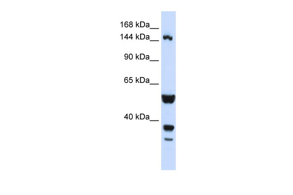
Human HeLa; WB Suggested Anti-ABI3BP Antibody Titration: 0.2-1 ug/ml. ELISA Titer: 1:62500. Positive Control: Hela cell lysate; ABI3BP antibody - N-terminal region (ARP47157_P050) in Human HeLa cells using Western Blot
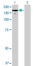
Western Blot analysis of ABI3BP expression in transfected 293T cell line (H00025890-T01) by ABI3BP MaxPab polyclonal antibody.Lane 1: ABI3BP transfected lysate(118.25 KDa).Lane 2: Non-transfected lysate.
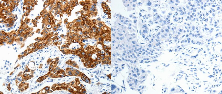
Predicted cell location: Secreted. Positive control: Human lung and ovarian cancer tissue . Recommended dilution: 1/25-100 The image on the left is immunohistochemistry of paraffin-embedded human lung cancer tissue using ABI3BP antibody at dilution 1/25, on the right is treated with the synthetic peptide. (Original magnification:x 200)

Predicted cell location: Secreted. Positive control: Human lung and ovarian cancer tissue . Recommended dilution: 1/25-100 The image on the left is immunohistochemistry of paraffin-embedded human ovarian cancer tissue using ABI3BP antibody at dilution 1/25, on the right is treated with the synthetic peptide. (Original magnification:x200)
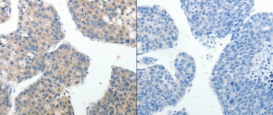
Predicted cell location: Secreted. Positive control: Human liver and gastric cancer tissue . Recommended dilution: 1/15-50 The image on the left is immunohistochemistry of paraffin-embedded human liver cancer tissue using ABI3BP antibody at dilution 1/20, on the right is treated with the synthetic peptide. (Original magnification:x 200)
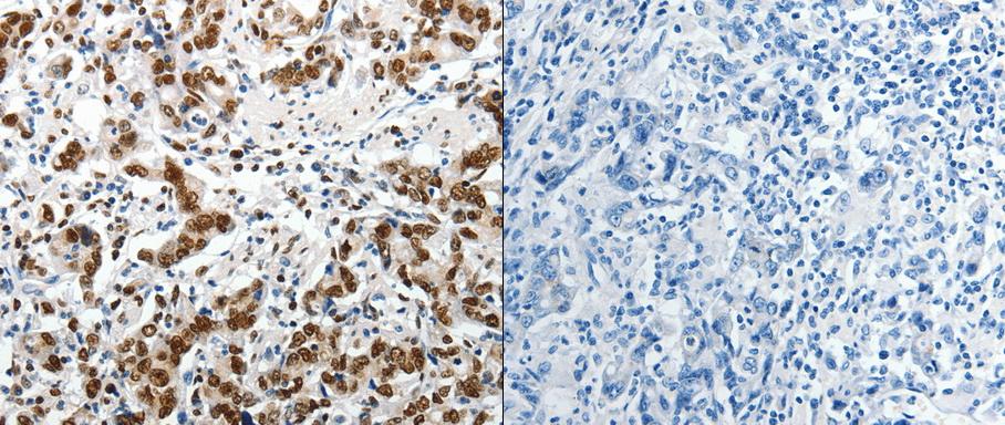
Predicted cell location: Secreted. Positive control: Human liver and gastric cancer tissue . Recommended dilution: 1/15-50 The image on the left is immunohistochemistry of paraffin-embedded human gastric cancer tissue using ABI3BP antibody at dilution 1/20, on the right is treated with the synthetic peptide. (Original magnification:x200)
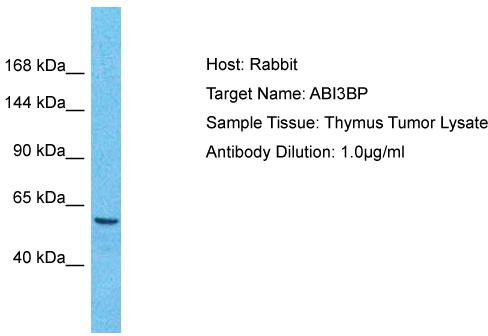
Host: Rabbit; Target Name: ABI3BP; Sample Tissue: Thymus Tumor lysates; Antibody Dilution: 1.0 ug/ml
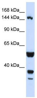
WB Suggested Anti-ABI3BP Antibody Titration: 0.2-1 ug/ml; ELISA Titer: 1: 62500; Positive Control: Hela cell lysate
Primary Antibodies
| Catalog No. | Host | Iso. | Clone | Pres. | React. | Applications | |
|---|---|---|---|---|---|---|---|
| TA322351 | ABI3BP antibody |
||||||
| Anti-ABI3BP Rabbit Polyclonal Antibody | |||||||
 |
Rabbit | IgG | Purified | Hu, Ms, Rt | E, P |
0.1 ml /
€335.00
|
|
| OriGene Technologies, Inc. | |||||||
 +1 additional image +1 additional image |
|||||||
| TA322352 | ABI3BP antibody |
||||||
| Anti-ABI3BP Rabbit Polyclonal Antibody | |||||||
 |
Rabbit | IgG | Purified | Hu, Ms, Rt | E, P |
0.1 ml /
€335.00
|
|
| OriGene Technologies, Inc. | |||||||
| +1 additional image |
|||||||