APH1A antibody
Principal name
APH1A antibody
Alternative names for APH1A antibody
Gamma-secretase subunit APH-1A, APH-1A, Aph-1 alpha, PSF, Presenilin-stabilization factor, CGI-78, aph-1
Ncbi ID
NP_001071096.1, 9606, NM_001077628, AAH08732, NP_666216.1, NM_146104, NM_016022
Available reactivities
Available hosts
Available applications
Western blot / Immunoblot (WB), Paraffin Sections (P), Enzyme Immunoassay (E)
Background of APH1A antibody
- Kit Component:
- - KN301394G1, Aph1a gRNA vector 1 in pCas-Guide vector
- KN301394G2, Aph1a gRNA vector 2 in pCas-Guide vector
- KN301394D, donor vector containing left and right homologous arms and Luciferase-Puro functional cassette
- GE100003, scramble sequence in pCas-Guide vector - CRISPR Manual:
- CRISPR/CAS9 Genome Editing Manual
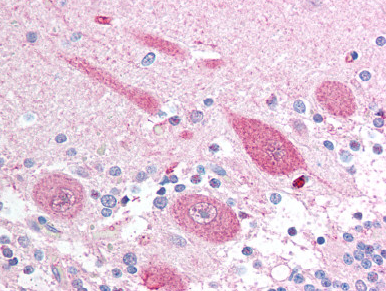
Brain, cerebellum: Formalin-Fixed Paraffin-Embedded (FFPE)
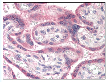
Figure 1. Formalin-Fixed Paraffin-Embedded (FFPE) on Placenta.

Western blot analysis of APH1A polyclonal antibody ( Cat # PAB4887 ) in A2058 cell lysate ( 35 µg/lane ) . APH1A ( arrow ) was detected using the purified polyclonal antibody.

APH1A polyclonal antibody ( Cat # PAB7304 ) ( 0.05 µg/ml ) staining of human brain ( cerebral cortex ) lysate ( 35 µg protein in RIPA buffer ) . Primary incubation was 1 hour. Detected by chemiluminescence.
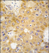
Formalin-fixed and paraffin-embedded human hepatocarcinoma tissue reacted with APH1 antibody (N-term), which was peroxidase-conjugated to the secondary antibody, followed by DAB staining.
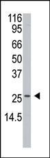
Western blot analysis of anti-APH1 Antibody (N-term) in A2058 cell line lysates (35ug/lane). APH1 (arrow) was detected using the purified Pab.
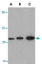
Western blot analysis of APH1A in RAW264.7 cell lysate with APH1A polyclonal antibody ( Cat # PAB13237 ) at ( A ) 0.5, ( B ) 1 and ( C ) 2 µg/ml.
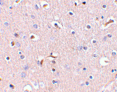
Immunohistochemistry of APH1A in human brain tissue with APH1A polyclonal antibody ( Cat # PAB13238 ) at 5 µg/ml.
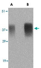
Western blot analysis of APH1A in human brain tissue lysate with APH1A polyclonal antibody ( Cat # PAB13238 ) at ( A ) 0.5 and ( B ) 1 µg/ml.
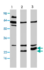
Western blot analysis of extract from Neuro 2a cells, using APH1A polyclonal antibody ( Cat # PAB8483 ) . Lane 1: mock, lane 2: APH1A ( 247 a.a. ) and lane 3: APH1A ( 265 a. a. ) .
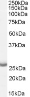
TA303251 (0.05µg/ml) staining of Human Brain (Cerebral Cortex) lysate (35µg protein in RIPA buffer). Primary incubation was 1 hour. Detected by chemiluminescence.
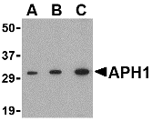
Western blot analysis of APH1 in RAW264.7 cell lysate with APH1 antibody at (A) 0.5, (B) 1 and (C) 2 µg/ml.
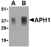
Western blot analysis of APH1 in human brain tissue lysate with APH1 antibody at (A) 0.5 and (B) 1 ug/ml.

Immunohistochemistry of APH1 in human brain tissue with APH1 antibody at 5 ug/ml.
Primary Antibodies
| Catalog No. | Host | Iso. | Clone | Pres. | React. | Applications | |
|---|---|---|---|---|---|---|---|
| TA303251 | APH1A antibody |
||||||
| Goat Polyclonal Antibody against APH1A | |||||||
 |
Goat | Aff - Purified | Hu, Ms, Rt | WB |
0.1 mg /
€360.00
|
||
| OriGene Technologies, Inc. | |||||||
| TA306377 | APH1A (N-term) antibody |
||||||
| Rabbit Polyclonal APH1 Antibody | |||||||
 |
Rabbit | IgG | Aff - Purified | Hu, Ms | E, WB |
0.1 mg /
€415.00
|
|
| OriGene Technologies, Inc. | |||||||
| TA306378 | APH1A (Center) antibody |
||||||
| Rabbit Polyclonal APH1 Antibody | |||||||
 |
Rabbit | IgG | Aff - Purified | Hu, Ms, Rt | E, P, WB |
0.1 mg /
€415.00
|
|
| OriGene Technologies, Inc. | |||||||
 +1 additional image +1 additional image |
|||||||
| AP07271PU-N | APH1A antibody |
||||||
 |
Rabbit | Aff - Purified | Hu, Ms, Rt | P, WB |
50 µg /
€425.00
|
||
| OriGene Technologies GmbH | |||||||
| AP07656PU-N | APH1A antibody |
||||||
 |
Rabbit | Purified | Hu, Ms | P, WB |
50 µg /
€425.00
|
||
| OriGene Technologies GmbH | |||||||
| AP13314PU-N | APH1A (N-term) antibody |
||||||
 |
Rabbit | Ig | Purified | Hu | P, WB |
0.4 ml /
€415.00
|
|
| OriGene Technologies GmbH | |||||||
| +1 additional image |
|||||||
Lysates
| Catalog No. | ||
|---|---|---|
| LY421466 | APH1A overexpression lysate |
|
 |
0.1 mg /
€295.00
|
|
| OriGene Technologies, Inc. |