ARF1 antibody
Principal name
ARF1 antibody
Alternative names for ARF1 antibody
ADP-ribosylation factor 1
SwissProt ID
P36397 (Arath), P51821 (Chlre), P84077 (Human), P84078 (Mouse), P84079 (Rat), P84080 (Bovin)
Ncbi ID
9606, NP_182239.1, NP_001019397, NP_071963, NP_001649, NM_001658, AAH11358.1, NP_001019398, NP_001019397.1, NP_001123880.1, NM_022518, NM_001024227, NM_001024228, NM_001024226
Available reactivities
Available hosts
Available applications
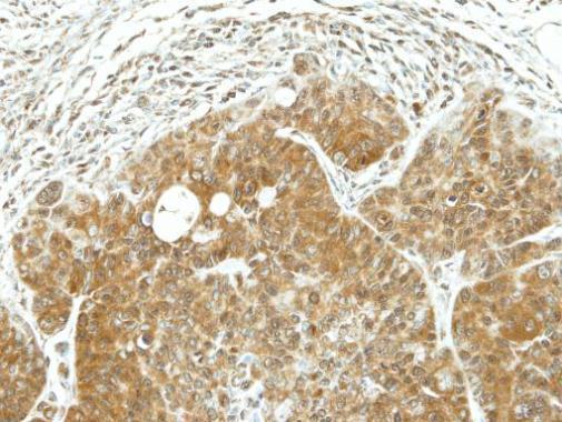
Enzyme Immunoassay (E), Western blot / Immunoblot (WB), Flow Cytometry (F), Immunocytochemistry/Immunofluorescence (ICC/IF), Paraffin Sections (P)
Background of ARF1 antibody
Ras superfamily of small GTPases regulates a wide variety of cellular processes including intracellular trafficking of proteins. The Ras proteins acts as molecular switches by cycling between inactive GDP-bound and active GTP-bound state. Guanine exchange factors (GEFs) Among the various Ras proteins, the ADP-ribosylation factor proteins (ARFs) are well characterized regulators of vesicle formation in intracellular membrane traffic while Rho proteins regulate the actin cytoskeleton (1). The Arf family members have unique structural device, called the ‘interswitch toggle’, that implements communication between the nucleotide-binding site and a unique N-terminal amphipathic helix that mediates the interaction of Arf proteins with membranes, these structural motifs distinguish them from other small GTPases (2). When GDP is bound to ARF proteins the amphipathic helix is positioned in a hydrophobic pocket, whereas in the GTP-bound forms it is displaced by the interswitch toggle and stabilizes interaction of the protein with membrane lipid bilayer. Thus, the GTP-GDP cycle is coupled to membrane-cytosol cycle and thereby ARF proteins are able to regulate vesicular traffic and organelle structure formation (2).
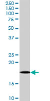
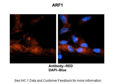
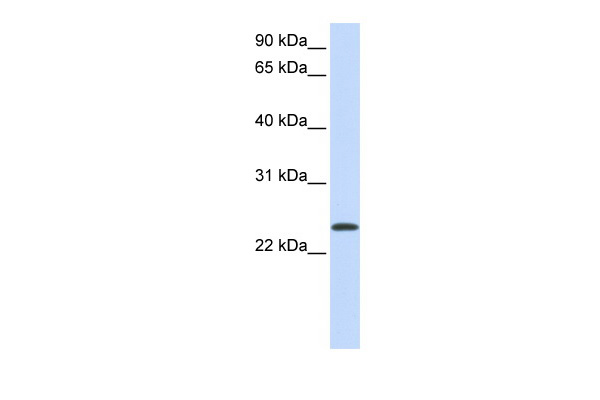
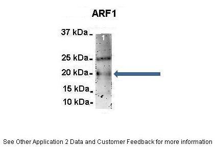
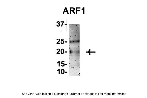
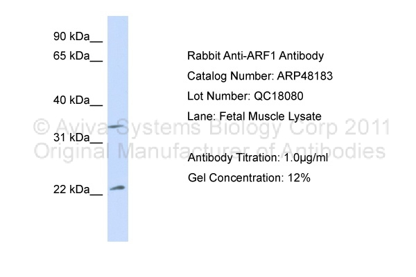
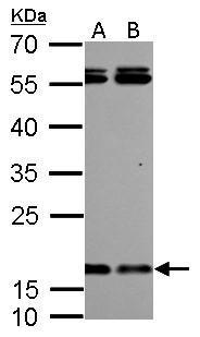
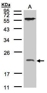
The mammalian ARF family comprise of 5 members (ARF1-6), there is no ARF2. ARF1 and ARF6 are most abundantly expressed and located in distinct cellular locations at Golgi complex and early-endosome-plasma membrane interface and are most distantly related with only 67% sequence identity. ARF1 is mostly known for its role in vesicle budding and Golgi regulation through the recruitment of coat proteins and in the regulation of lipid-modifying enzymes and actin organizing components (3). The crystal structure of ARF-BD (ARF binding domain) comprise of a PH domain adjacent to a C-terminal helix and ARF interacts with these two regions via its “interswitch toggle” (4). ARF1 protein has 188 amino acids and is expressed in at least 3 variant forms, ARF1 antibodies will label all variants equally. The deactivation of ARF1 induces redistribution of entire Golgi complex to ER, the GTP bound form triggers the association with coat protein with the Golgi membranes. Purified ARF proteins do not have GTPase activity suggesting that GTP hydrolysis is dependent on GTPase activating protein (GAP) a 49kDa protein (4). The GTP bound ARF1 and 3 proteins also interact with several interacting protein (ARFGAPs) including protein interacting with C kinase 1 (PICK1) to mediate their cellular processes.
General readings
1. Hall A (2005) Rho GTPases and the control of cell behavior. Biochem Soc Trans 33: 891–895
2. Antonny B, Beraud-Dufour S, Chardin P, Chabre M (1997) Biochemistry 36: 4675–4684
3. D'Souza-Schorey C, Chavrier P (2006) ARF proteins: roles in membrane traffic and beyond. Nat Rev Mol Cell Biol 7: 347–358
4. Julie Ménétrey, Mylène Perderiset,Jérome Cicolari, Thierry Dubois, Nadia Elkhatib, Fatima El Khadali, Michel Franco, Philippe Chavrier, and Anne Houdusse. EMBO J .26(7); Apr 4, 2007
5. Makler V, Cukierman E, Rotman M, Admon A, Cassel D. ADP-ribosylation factor-directed GTPase-activating protein. Purification and partial characterization. J Biol Chem. 1995 Mar
10;270(10):5232-7. Links


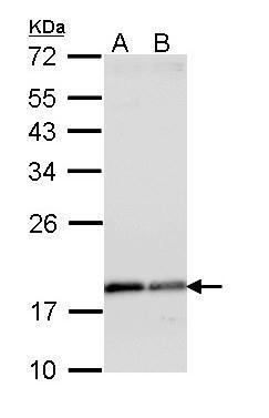
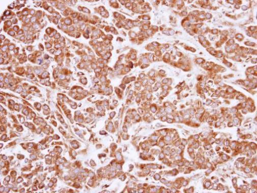
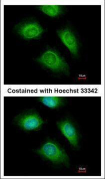
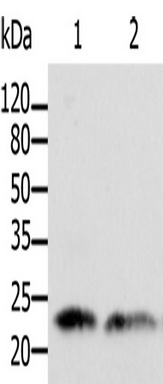
Primary Antibodies
| Catalog No. | Host | Iso. | Clone | Pres. | React. | Applications | |
|---|---|---|---|---|---|---|---|
| TA303176 | ARF1/2/3/4 (C-term) antibody |
||||||
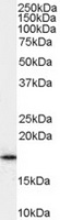
| Goat Polyclonal Antibody against ARF1 | |||||||
 |
Goat | Aff - Purified | Can, Hu, Ms, Rt | E, WB |
0.1 mg /
€360.00
|
||
| OriGene Technologies, Inc. | |||||||

