ARID1B antibody
Principal name
ARID1B antibody
Alternative names for ARID1B antibody
DAN15, KIAA1235, OSA2, Osa homolog 2, p250R, BRG1-associated factor 250b, AT-rich interactive domain-containing protein 1B, ARID domain-containing protein 1B, BRG1-binding protein hELD/OSA1
SwissProt ID
Q8NFD5 (Human)
Gene ID
57492 (ARID1B)
Ncbi ID
Available reactivities
Available hosts
Available applications
Enzyme Immunoassay (E), Immunocytochemistry/Immunofluorescence (ICC/IF), Western blot / Immunoblot (WB), Paraffin Sections (P)
Background of ARID1B antibody
Transient overexpression lysate of AT rich interactive domain 1B (SWI1-like) (ARID1B), transcript variant 2
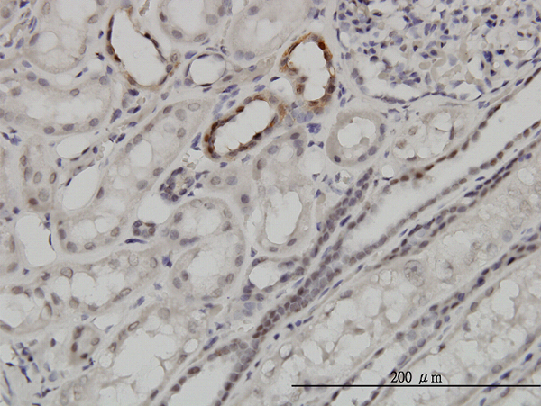
Immunoperoxidase of monoclonal antibody to ARID1B on formalin-fixed paraffin-embedded human kidney. [antibody concentration 3ug/ml]
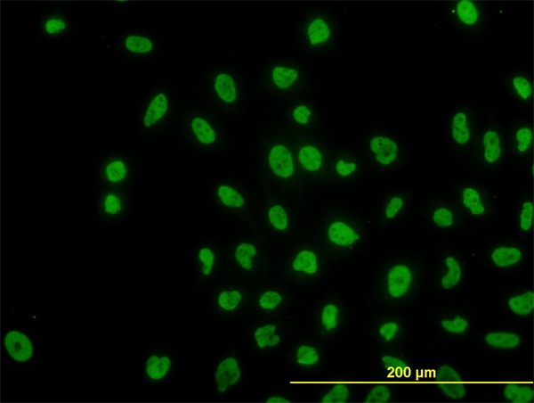
Immunofluorescence of monoclonal antibody to ARID1B on HeLa cell. [antibody concentration 10 ug/ml]
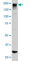
ARID1B monoclonal antibody (M01), clone 2D2 Western Blot analysis of ARID1B expression in HeLa NE ( Cat # L013V3 ).
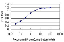
Detection limit for recombinant GST tagged ARID1B is approximately 0.03ng/ml as a capture antibody.
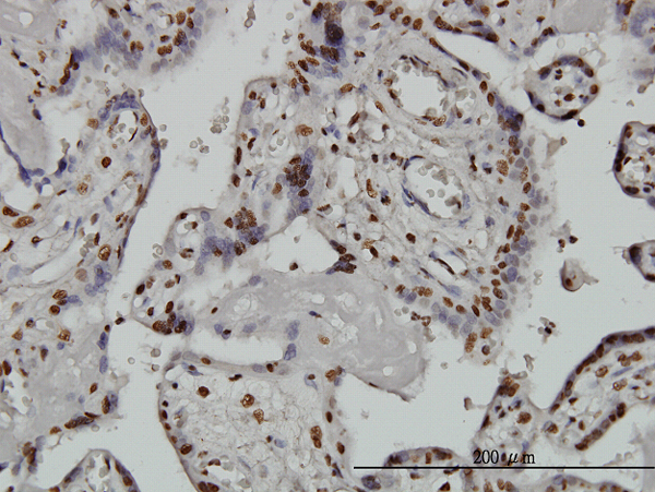
Immunoperoxidase of monoclonal antibody to ARID1B on formalin-fixed paraffin-embedded human placenta. [antibody concentration 3 ug/ml]
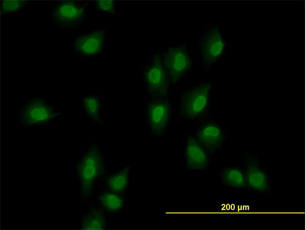
Immunofluorescence of monoclonal antibody to ARID1B on HeLa cell. [antibody concentration 10 ug/ml]

ARID1B monoclonal antibody (M02), clone 2F2 Western Blot analysis of ARID1B expression in Hela NE ( Cat # L013V3 ).
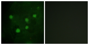

Immunohistochemical analysis of BAF250B staining in human breast cancer formalin fixed paraffin embedded tissue section. The section was pre-treated using heat mediated antigen retrieval with sodium citrate buffer (pH 6.0). The section was then incubated with the antibody at room temperature and detected using an HRP conjugated compact polymer system. DAB was used as the chromogen. The section was then counterstained with haematoxylin and mounted with DPX.
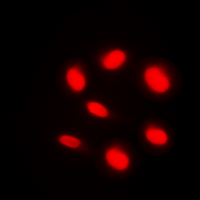
Immunofluorescent analysis of BAF250B staining in HeLa cells. Formalin-fixed cells were permeabilized with 0.1% Triton X-100 in TBS for 5-10 minutes and blocked with 3% BSA-PBS for 30 minutes at room temperature. Cells were probed with the primary antibody in 3% BSA-PBS and incubated overnight at 4 C in a humidified chamber. Cells were washed with PBST and incubated with a DyLight 594-conjugated secondary antibody (red) in PBS at room temperature in the dark. DAPI was used to stain the cell nuclei (blue).
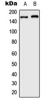
Western blot analysis of BAF250B expression in HeLa (A); HepG2 (B) whole cell lysates.
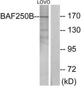
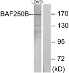
Western blot analysis of extracts from LOVO cells, using BAF250B antibody.The lane on the right is treated with the synthesized peptide.
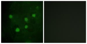
Immunofluorescence analysis of HUVEC cells, using BAF250B antibody.The picture on the right is treated with the synthesized peptide.
Primary Antibodies
| Catalog No. | Host | Iso. | Clone | Pres. | React. | Applications | |
|---|---|---|---|---|---|---|---|
| TA315663 | ARID1B antibody |
||||||
| Rabbit polyclonal anti-BAF250B antibody | |||||||
 |
Rabbit | IgG | Purified | Hu, Rt | E, ICC/IF, WB |
0.1 ml /
€335.00
|
|
| OriGene Technologies, Inc. | |||||||
 +1 additional image +1 additional image |
|||||||
Lysates
| Catalog No. | ||
|---|---|---|
| LY412359 | ARID1B overexpression lysate |
|
 |
0.1 mg /
€495.00
|
|
| OriGene Technologies, Inc. |
Human Lenti ORF Particles
| Catalog No. | ||
|---|---|---|
| RC214830L1V | Lenti ORF particles, ARID1B (Myc-DDK tagged) - Human AT rich interactive domain 1B (SWI1-like) (ARID1B), transcript variant 2, 200 uL, >10^7 TU/mL |
|
|
200 µl /
€2,420.00
|
|
| OriGene Technologies, Inc. | ||