Bcl-2 antibody
Principal name
Bcl-2 antibody
Alternative names for Bcl-2 antibody
BCL2, Bcl-2 alpha, Apoptosis regulator Bcl-2, B-Cell CLL/lymphoma 2
SwissProt ID
O02718 (Bovin), P10415 (Human), P10417 (Mouse), P49950 (Rat), Q00709 (Chick), Q6R755 (Canfa)
Ncbi ID
NP_000624, NP_000624.2, 9606, 10090, NP_058689.1, AAH27258, NM_009741, NM_177410, NM_016993, NM_000633
Available reactivities
Available hosts
Available applications
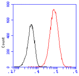
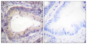
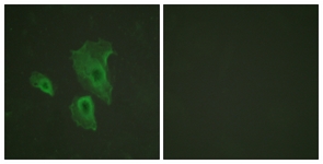
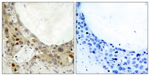
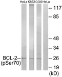
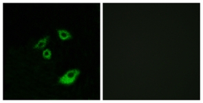
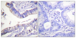
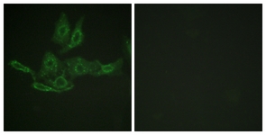
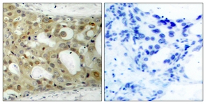
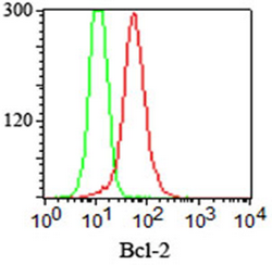
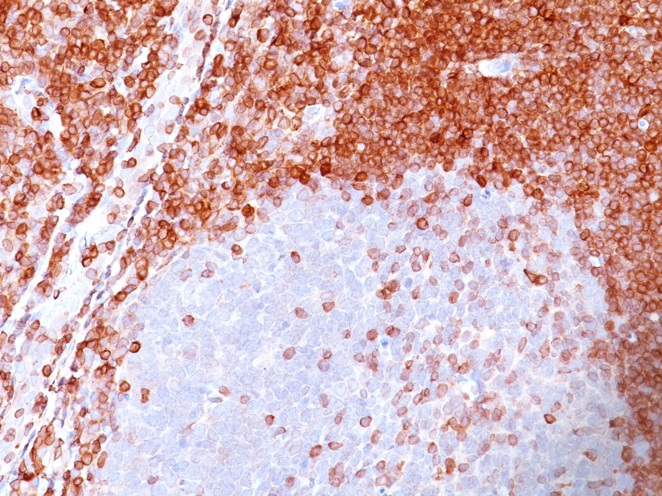
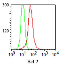
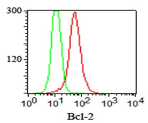
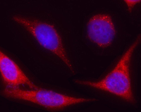
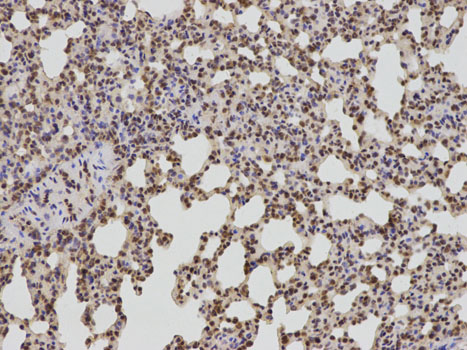
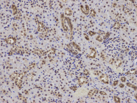
Western blot / Immunoblot (WB), Paraffin Sections (P), Frozen Sections (C), Immunoprecipitation (IP), Flow Cytometry (F), Enzyme Immunoassay (E), Immunocytochemistry/Immunofluorescence (ICC/IF), Neutralisation (NEUT)
Background of Bcl-2 antibody
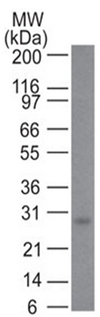
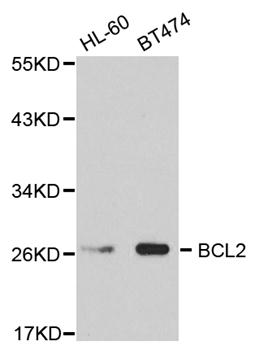
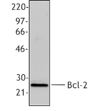
Bcl-2 is a 26 kDa member of the family of proteins involved in regulation of programmed cell death, or apoptosis. It is expressed in a variety of both normal and neoplastic tissues and appears to be membranebound since it is detectable in crude membrane and nuclear fractions, but not in the soluble fraction of cell lysates. Overexpression of Bcl-2 can prevent apoptosis, while formation of heterodimers with Bax, another Bcl-2-related protein, inhibits Bcl-2’s ability to promote cell survival.
Apoptosis, or programmed cell death, is a well-documented phenomenon in many cellular systems. (Ref.2) It plays a key role in tissue and organ development as well as in adult tissues during cell turnover. Apoptosis can be induced by a variety of internal and external stimuli including growth factor deprivation, cytokine treatment, antigen-receptor engagement, cell-cell interactions, irradiation and glucocorticoid treatment. (Ref.3)
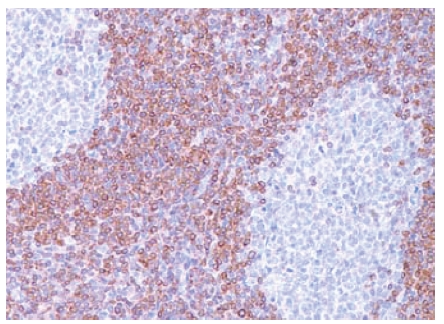
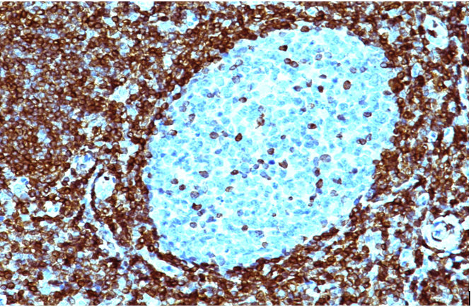
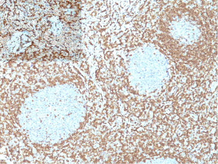
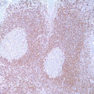
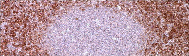
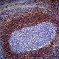
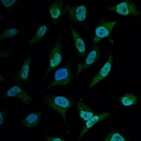
Bcl-2 is a widely studied protein that has been shown to be a potent inhibitor of programmed cell death. It has been localized to the outer mitochondrial membrane, perinuclear membrane, and endoplasmic reticulum. Bcl-2 is expressed in memory and resting, or other long-lived lymphoid cells, follicular mantle cells, medullary thymocytes, and lymphomas. Germical center cells and cortical thymocytes are negative for Bcl-2. Upregulation of Bcl-2 prevents or delays apoptosis induced by a variety of stimuli, including growth factor deprivation, gamma-irradiation, glucocorticoids, and chemotherapeutic agents. During lymphoid development, expression of the Bcl-2 protein appears to be regulated in a stage-specific manner, and is thought to be a survival signal for positive selection.
General readings
1. Bartoszewski R et al. Mangiferin has an additive effect on the apoptotic properties of hesperidin in Cyclopia sp. tea extracts. PLoS One 9:e92128 (2014). WB ; Human .
2. Adams SM et al. Soy isoflavones genistein and daidzein exert anti-apoptotic actions via a selective ER-mediated mechanism in neurons following HIV-1 Tat(1-86) exposure. PLoS One 7:e37540 (2012). ELISA ; Rat .
3. Fujita H et al. Novel method for measuring active tension generation by C2C12 myotube using UV-crosslinked collagen film. Biotechnol Bioeng 106:482-9 (2010). WB ; Mouse .
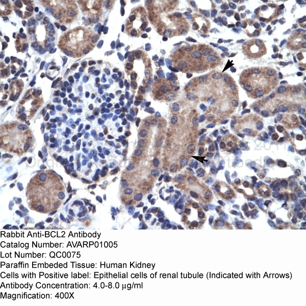
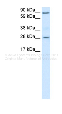


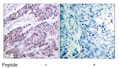
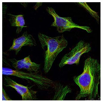
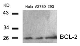
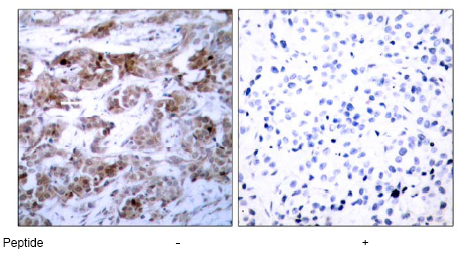
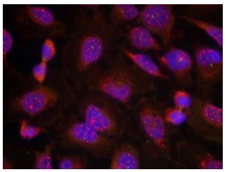
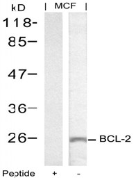
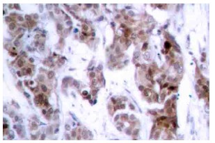

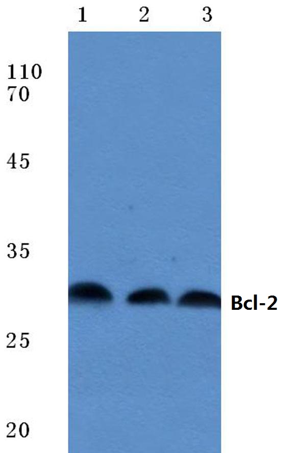
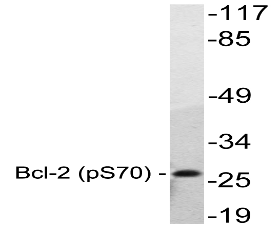
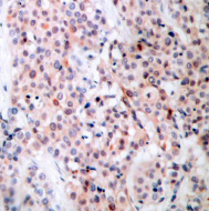
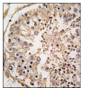
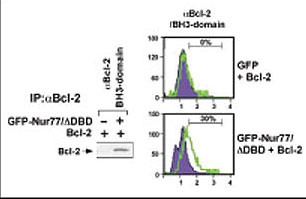
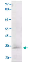
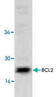
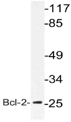
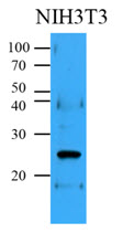
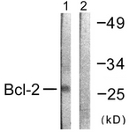
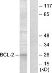
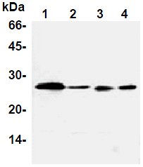
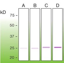
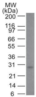
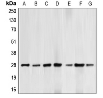
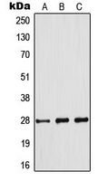
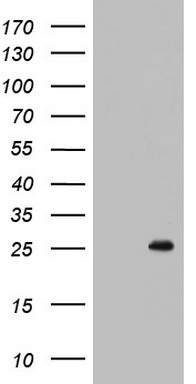
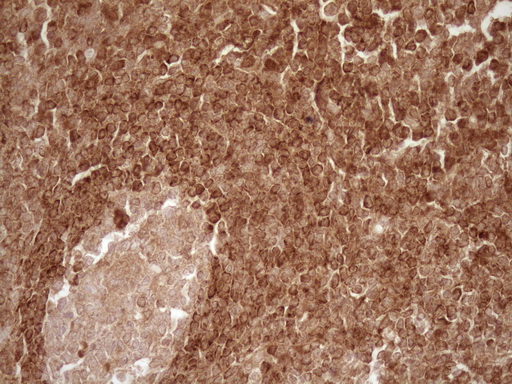
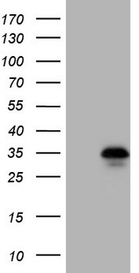
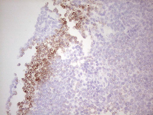
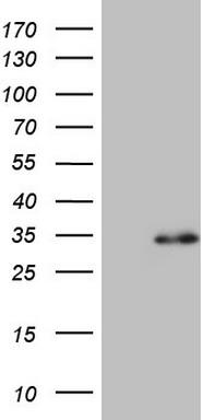
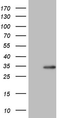
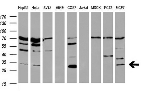
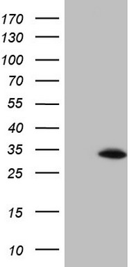
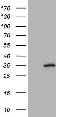
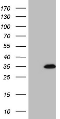
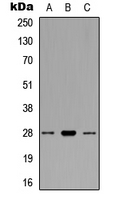
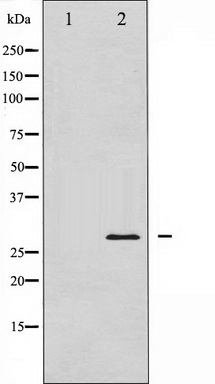
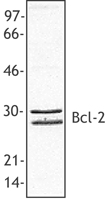
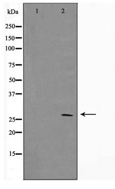
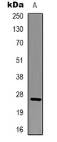
Lane1:MCF-7 cell lysate
Lane2:NIH-3T3 cell lysate
Lane3:Raw264.7 cell lysate
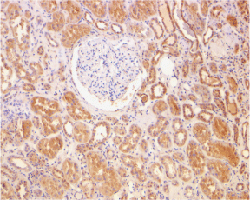
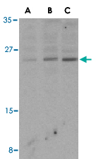
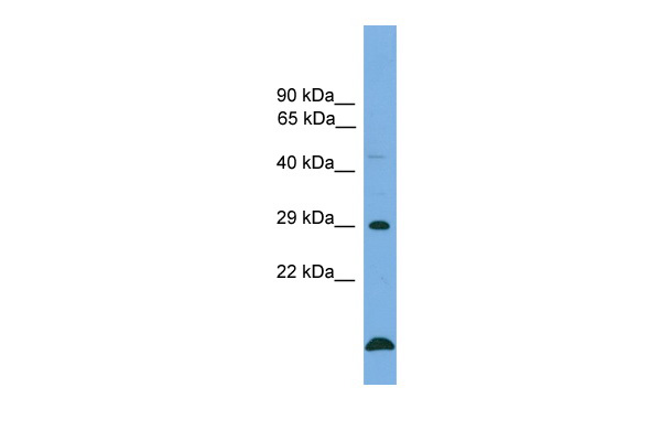
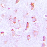
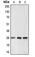
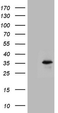
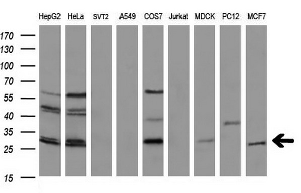
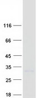
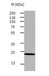
Lane1: MCF-7 whole cell lysate treated with PMA
Lane2: Mouse brain tissue lysate
Lane3: Rat kidney tissue lysate


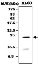

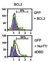
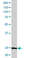


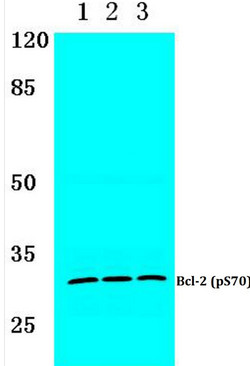
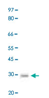
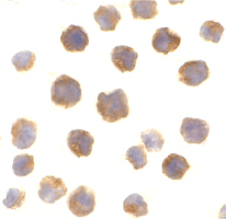
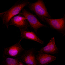
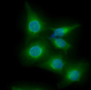
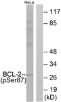
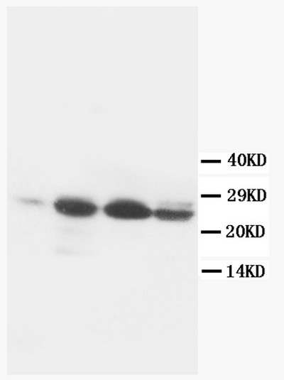
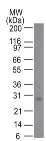
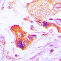
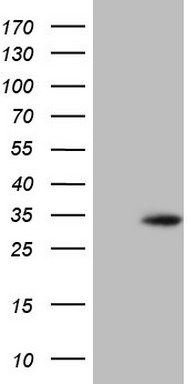
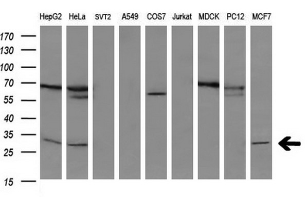
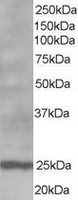
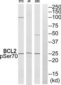
Lane 1: Hela cell lysate treated with colchicine.
Lane 2: Raw264.7 cell lysate treated with colchicine.
Lane 3: PC12 cell lysate treated with colchicine.















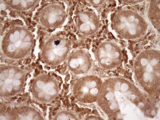
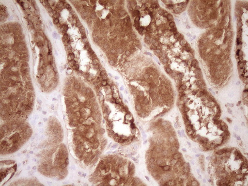
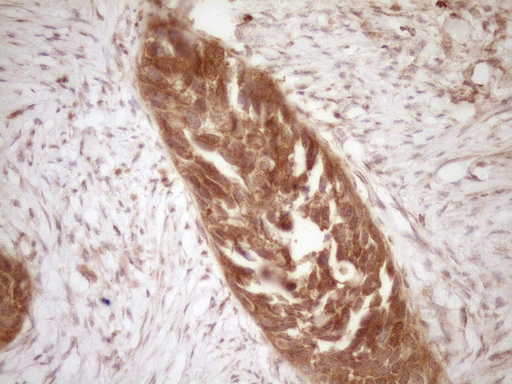

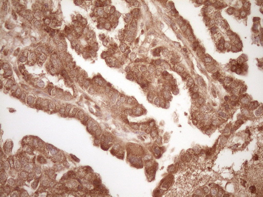
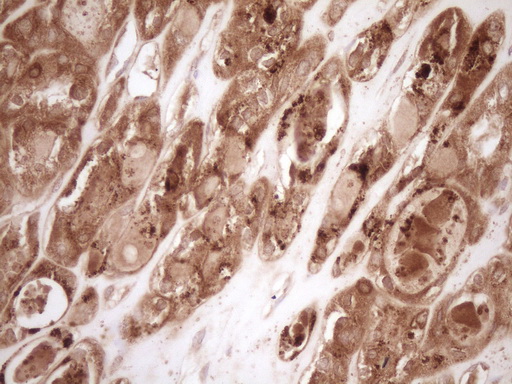
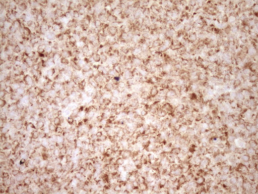
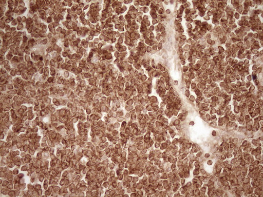
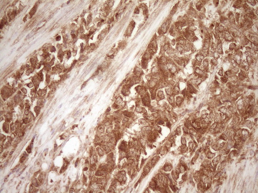






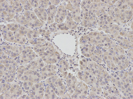
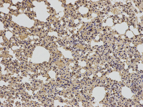
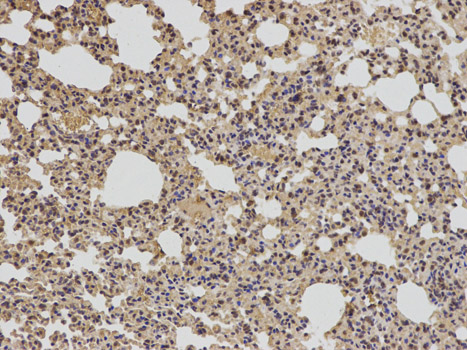
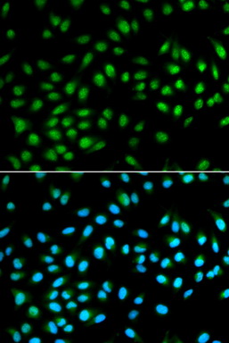
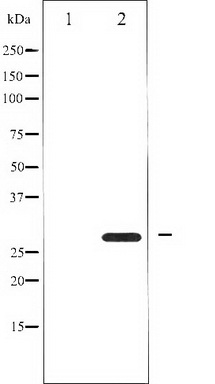
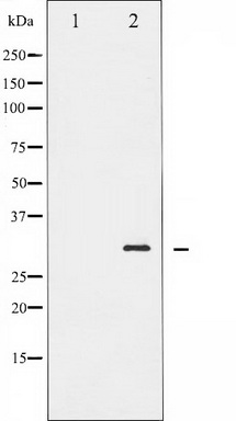
Primary Antibodies
| Catalog No. | Host | Iso. | Clone | Pres. | React. | Applications | |
|---|---|---|---|---|---|---|---|
| AM10050SU-N | Bcl-2 antibody |
||||||
 |
Mouse | IgG1 | 124 | Supernatant | Hu | P, WB |
1 ml /
€450.00
|
| OriGene Technologies GmbH | |||||||
| AM32275SU-N | Bcl-2 antibody |
||||||
 |
Rabbit | IgG | E17 | Supernatant | Hu | C, P |
1 ml /
€820.00
|
| OriGene Technologies GmbH | |||||||