BVES antibody
Principal name
BVES antibody
Alternative names for BVES antibody
POP1, POPDC1, Blood vessel epicardial substance
Ncbi ID
9606, NP_009004, NP_001186492, NP_001186492.1, NP_077247.1, NM_147147, NM_007073
Available reactivities
Available hosts
Available applications
Enzyme Immunoassay (E), Western blot / Immunoblot (WB)
Background of BVES antibody
- Kit Component:
- - KN302326G1, Bves gRNA vector 1 in pCas-Guide vector
- KN302326G2, Bves gRNA vector 2 in pCas-Guide vector
- KN302326D, donor vector containing left and right homologous arms and Luciferase-Puro functional cassette
- GE100003, scramble sequence in pCas-Guide vector - CRISPR Manual:
- CRISPR/CAS9 Genome Editing Manual

BVES monoclonal antibody (M02), clone 3F7. Western Blot analysis of BVES expression in PC-12 ( Cat # L012V1 ).
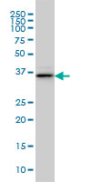
BVES monoclonal antibody (M02), clone 3F7 Western Blot analysis of BVES expression in IMR-32 ( Cat # L008V1 ).
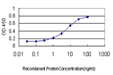
Detection limit for recombinant GST tagged BVES is approximately 0.1ng/ml as a capture antibody.
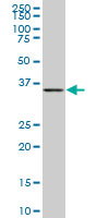
BVES monoclonal antibody (M01), clone 1C3. Western Blot analysis of BVES expression in IMR-32 ( Cat # L008V1 ).
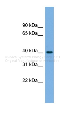
Human Stomach; WB Suggested Anti-BVES Antibody Titration: 0.2-1 ug/ml. Positive Control: Human Stomach; BVES antibody - middle region (ARP46823_P050) in Human Stomach cells using Western Blot
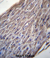
BVES Antibody (C-term) (Cat. #AP50411PU-N)immunohistochemistry analysis in formalin fixed and paraffin embedded human heart tissue followed by peroxidase conjugation of the secondary antibody and DAB staining.This data demonstrates the use of BVES Antibody (C-term) for immunohistochemistry. Clinical relevance has not been evaluated.
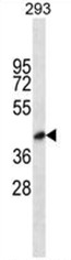
BVES Antibody (C-term) (Cat. #AP50411PU-N) western blot analysis in 293 cell line lysates (35µg/lane).This demonstrates the BVES antibody detected the BVES protein (arrow).
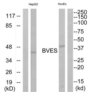
Western blot analysis of extracts from HepG2 cells and HuvEc cells, using BVES antibody.The lane on the right is treated with the synthesized peptide.
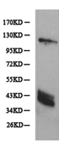
Predicted band size: 41 kDa . Positive control: Skeletal muscle tissue lysate. Recommended dilution: 1/500-2000. (Gel: 10%SDS-PAGE Lysate: 40 ug Primary antibody: 1/500 dilution Secondary antibody: Goat anti Rabbit IgG - H&L (HRP) at 1/10000 dilution Exposure time: 1 minute)
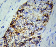
Predicted cell location: Cytoplasm. Positive control: Ovary tumor tissue. Recommended dilution: 1/50-200 The image is immunohistochemistry of paraffin-embedded Ovary tumor tissue using BVES antibody at dilution 1/50

Western blot analysis of extracts of various cell lines, using BVES antibody.
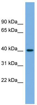
WB Suggested Anti-BVES Antibody Titration: 0.2-1 ug/ml; Positive Control: Human Stomach
Primary Antibodies
| Catalog No. | Host | Iso. | Clone | Pres. | React. | Applications | |
|---|---|---|---|---|---|---|---|
| TA315155 | BVES antibody |
||||||
| Rabbit polyclonal anti-BVES antibody | |||||||
 |
Rabbit | IgG | Purified | Hu, Ms, Rt | E, WB |
0.1 ml /
€335.00
|
|
| OriGene Technologies, Inc. | |||||||
| TA324122 | BVES antibody |
||||||
| Anti-BVES Rabbit Polyclonal Antibody | |||||||
 |
Rabbit | IgG | Purified | Hu, Ms, Rt | WB |
0.1 ml /
€335.00
|
|
| OriGene Technologies, Inc. | |||||||
 +1 additional image +1 additional image |
|||||||
| TA326775 | BVES antibody |
||||||
| BVES Rabbit Polyclonal Antibody | |||||||
 |
Rabbit | IgG | Purified | Hu, Ms, Rt | WB |
0.1 mg /
€335.00
|
|
| OriGene Technologies, Inc. | |||||||
| AP50411PU-N | BVES (C-term) antibody |
||||||
 |
Rabbit | Ig | Aff - Purified | Hu | WB |
0.4 ml /
€415.00
|
|
| OriGene Technologies GmbH | |||||||
| +1 additional image |
|||||||