CD19 antibody
Principal name
CD19 antibody
Alternative names for CD19 antibody
Leu-12, B-cell marker, Differentiation antigen CD19, B-lymphocyte surface antigen B4
Ncbi ID
NP_001171569.1, NP_033974.2, 9606, NP_001171569, NP_033974, NP_001761, NM_001770, NM_001178098
Available reactivities
Available hosts
Available applications
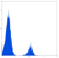
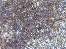
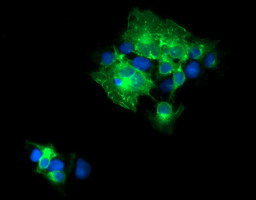
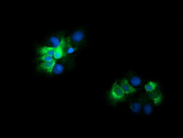
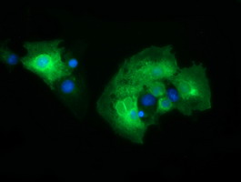
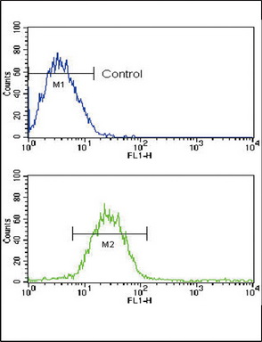
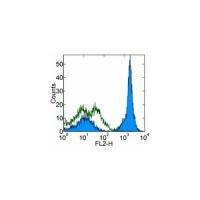
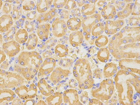
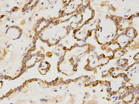
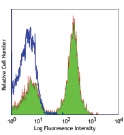
Frozen Sections (C), Functional assay (FN), Flow Cytometry (F), Immunoprecipitation (IP), Enzyme Immunoassay (E), Western blot / Immunoblot (WB), ELISA (detection) (E(detection)), Immunocytochemistry/Immunofluorescence (ICC/IF), Paraffin Sections (P), Luminex (LMNX)
Background of CD19 antibody
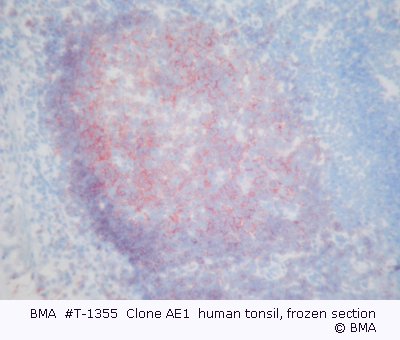
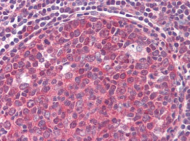
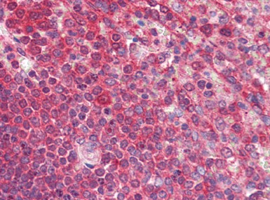
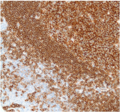
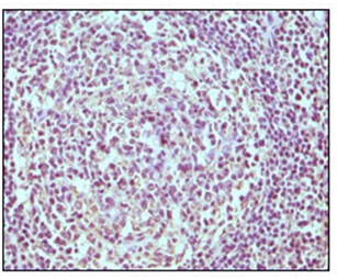
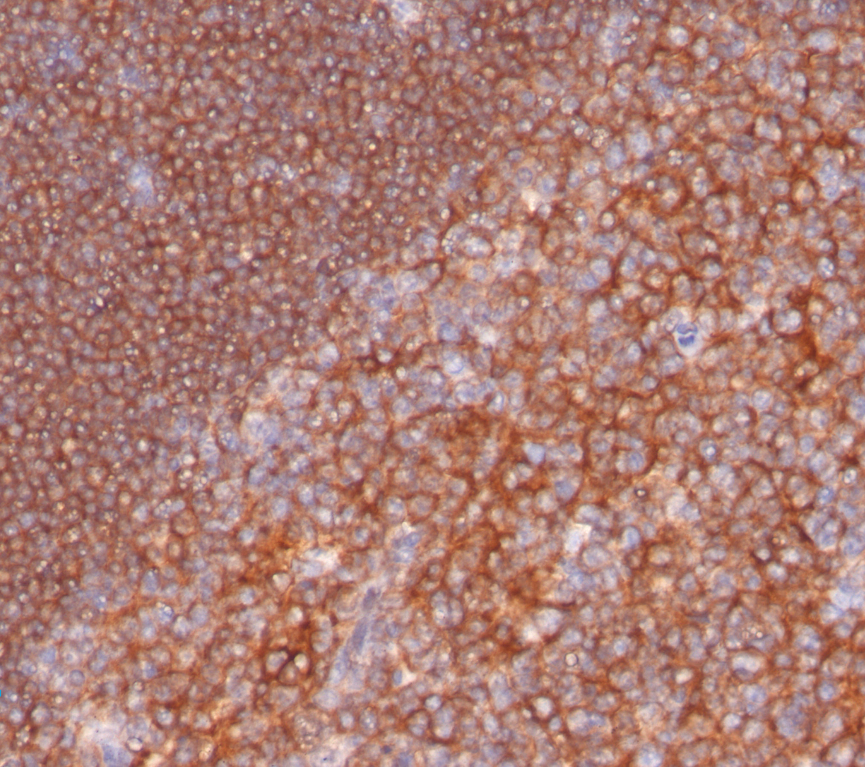
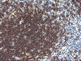
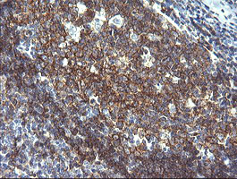
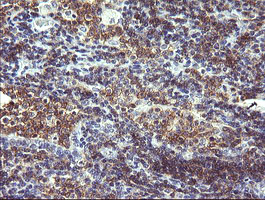
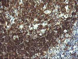
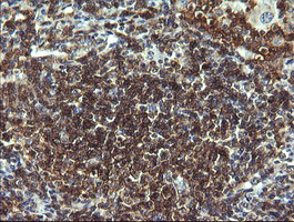
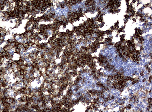
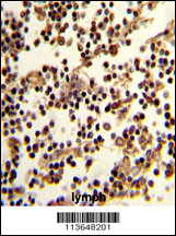
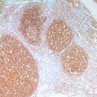
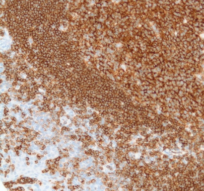
CD19 is present in both normal and malignant B cells and has long been considered to be the most reliable surface marker of this lineage over a wide range of maturational stages. In normal lymphoid tissue CD19 is observed in germinal centers (on both B cells and follicular dendritic cells), in mantle zone cells and in scattered cells in the interfollicular areas, with an overall immunoreactivity pattern similar to that of CD20 and CD22. However, in contrast to CD20, CD19 is also expressed in pre-B cells. CD19 has also been detected by flow cytometry in plasma cells isolated from human tissues. Recently, Masir et al. have described the expression of CD19 in normal lymphoid tissue and its loss in B-cell neoplasms. CD19 positivity is seen in the lymphoid follicle in germinal centers, the mantle zone, as well as in interfollicular T-cell areas including large cells with ‘dendritic’ morphology.
CD19 positivity is seen in the vast majority of B-cell neoplasms (B-lymphoblastic lymphoma, small lymphocytic lymphoma/CLL, mantle cell lymphoma, follicular lymphoma, Burkitt lymphoma, marginal zone lymphoma, diffuse large B-cell lymphoma, T-cell-rich B-cell lymphoma, lymphoblastic lymphoma, hairy cell leukaemia) and commonly at a lower intensity than normal B-cell elements. Plasma cell neoplasms are consistently negative as are T-cell neoplasms. In the Masir study, CD19 was undetectable in 14% of diffuse large B-cell lymphomas, 30% of T-cell-rich B-cell lymphomas and 75% of post-transplant B-lymphoproliferative disease. CD19 expression is not seen in Reed-Sternberg cells of classic Hodgkin’s disease.
General readings
1. Functional Properties of CD19+ B Lymphocytes Positively Selected from Buffy Coats by Immunomagnetic Separation. Funderud S., Erikstien B., Asheim H.C., Nustad K., Stokke T., Blomhoff H.K., Holte H, Smeland E.B., Eur. J. Immunol. 1990 Ja;20(1):201-6.
2. Thymic B Cells from Myasthenia Gravis Patients are Activated B Cells. Phenotypic and Functional Analysis. Leprince C., Cohen-Kaminsky S., Berrih-Aknin S., Vernet-Der Garabedian B., Treton D., Galanaud P., Richard Y., J. Immunol. 1990 Oct., 145(7):2115-22.
3. Prognostic Significance of CD34 Expression in Childhood B Precursor Acute Lymphocytic Leukemia: A Pediatric Onocology Group Study. Borowitz MJ, Shuster JJ, Civin CI, Carrol AJ, Look AT, Behm FG, Land VJ, Pullen DJ, Crist WM, J. Clin. Onol. 1990 Au;8(8):1389-98.
4. Biphenotypic Acute Leukemia in Adults. Sulak LE, Clare CN, Morale BA, Hansen KL, Montiel MM, Am. J. Clin. Path. 1990 Ju;94(1):54-8.
5. Intersection of the Complement and Immune Systems: A Signal Transduction Complex of the B Lymphocyte Containing Complement Receptor type 2 and CD19. Matsumoto AK, Kopicky-Burd J., Carter Rh, Tuveson DA, Tedder TF, Fearon, DT, J. Exp. Med. 1991 Jan. 173(1):55-64.
6. Immunofluorescence Measurement in a Flow Cytometer using Low-Power Helium Neon Laser Excitation. Shapiro, H.M, Glazer, A.N., Christenson, L., Williams, J.M., and Strom, T. B. Cytometry 4,276, 1983.
7. Comparison of Helium Neon and Dye lasers for Excitation of Allophycocyanin. Loken, M.R., Kiej, J.F. and Kelly, K.,A. Cytometry 8, 96, 1987.
Portions of this product are manufactured under license from Carnegie Mellon University, U.S. Patent Number 5,268,486.
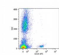
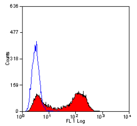
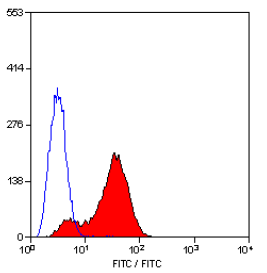
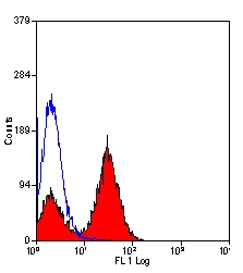
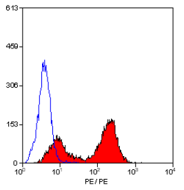

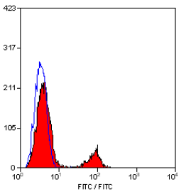

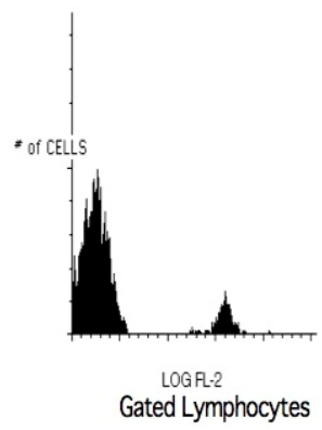

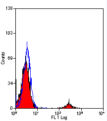
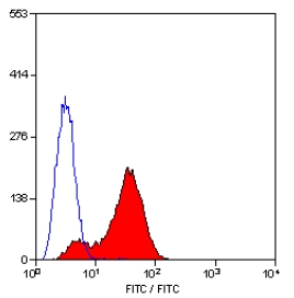

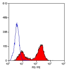
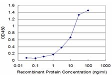
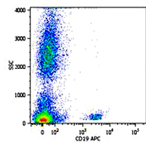

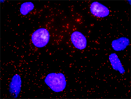

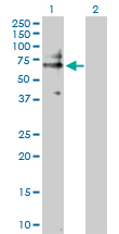
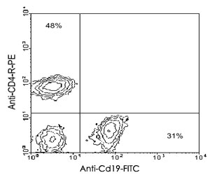
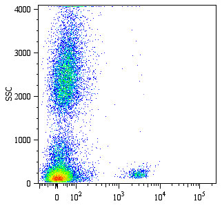
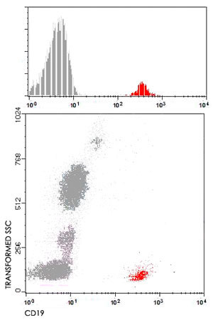
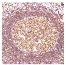
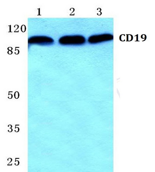
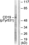
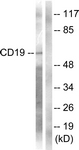
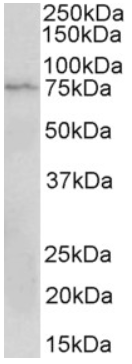
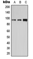
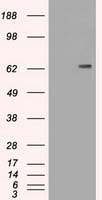
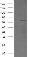
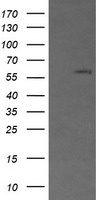
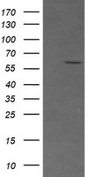
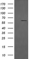
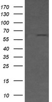
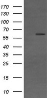
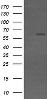
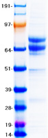
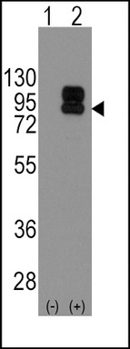
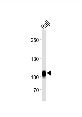
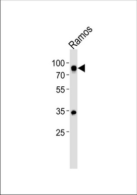
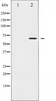
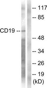
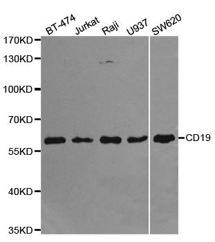
Lane 1: MCF-7 cell lysate.
Lane 2: Mouse spleen tissue lysate
Lane 3: Rat spleen tissue lysate.

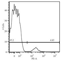





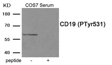
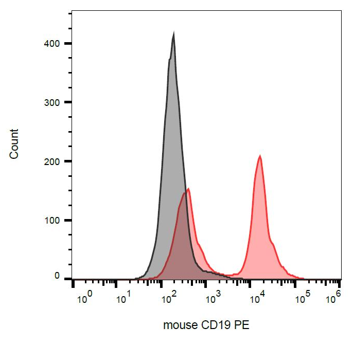
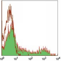





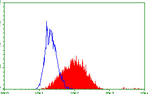
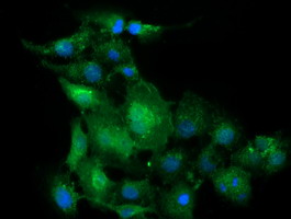
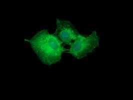






Primary Antibodies
| Catalog No. | Host | Iso. | Clone | Pres. | React. | Applications | |
|---|---|---|---|---|---|---|---|
| AM26049PU-N | CD19 antibody |
||||||
 |
Mouse | IgG1 | AE1 | Aff - Purified | Hu | C, F |
0.1 mg /
€310.00
|
| OriGene Technologies GmbH | |||||||