CEACAM7 antibody
Principal name
CEACAM7 antibody
Alternative names for CEACAM7 antibody
CGM2, Carcinoembryonic antigen-related cell adhesion molecule 7, Carcinoembryonic antigen CGM2
SwissProt ID
Q14002 (Human)
Gene ID
1087 (CEACAM7)
Ncbi ID
Available reactivities
Available hosts
Available applications
Enzyme Immunoassay (E), Frozen Sections (C), Immunocytochemistry/Immunofluorescence (ICC/IF), Flow Cytometry (F), Western blot / Immunoblot (WB)
Background of CEACAM7 antibody
- Kit Component:
- - KN212214G1, CEACAM7 gRNA vector 1 in pCas-Guide vector
- KN212214G2, CEACAM7 gRNA vector 2 in pCas-Guide vector
- KN212214D, donor vector containing left and right homologous arms and Luciferase-Puro functional cassette
- GE100003, scramble sequence in pCas-Guide vector - CRISPR Manual:
- CRISPR/CAS9 Genome Editing Manual
General readings
Thompson J, Seitz M, et al. (1997). Cancer Res. 57(9):1776-84.Kinugasa T, Kuroki M, et al. (1998). Int J Cancer. 76(1):148-53.
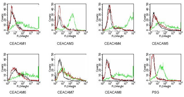
Figure 2. Members of the CEACAM family were expressed on BOSC cells after transient transfection withexpression vectors containing either the cDNA of CEACAM1, 5, 6, 7, 8 or a recombinant trans-membrane-anchored PSG1 fusion protein. Recognition of CEACAM3, CEACAM4 and PSG1 was tested on stably transfected HeLa (CEACAM3, PSG) and CHO cells (CEACAM4), respectively. Expression of the con-structs was confirmed with monoclonal antibodies known to recognize the corresponding proteins (CEACAM1, 3, 4, 5 and 6: D14HD11; CEACAM7: CAC2; CEACAM8: TET2; PSG: BAP1; green curves). An irrelevant monoclonal antibody served as a negative control (black curves). For specificity testing, protein G purified BAC2 was tested on all CEACAM transfectants. A positive signal was only obtained with CEACAM7 expressing cells (Red curves).
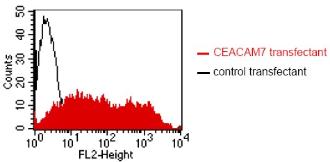
Figure 1. FACS analysis of BOSC23 cells using BAC2 Cat.DM1207. BOSC23 cells were transiently transfected with an expression vectorencoding either CEACAM7 (Red curve) or an irrelevant protein (control transfectant). Binding of BAC2 was detected with a PE conjugated secondary antibody. A positive signal was obtained only with CEACAM7 transfected cells.

Human Jurkat; WB Suggested Anti-CEACAM7 Antibody Titration: 0.2-1 ug/ml. ELISA Titer: 1:62500. Positive Control: Jurkat cell lysate; CEACAM7 antibody - N-terminal region (ARP60122_P050) in Human Jurkat cells using Western Blot
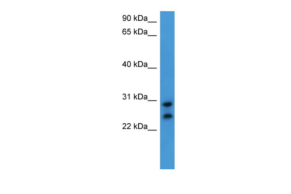
Human HeLa; WB Suggested Anti-CEACAM7 Antibody Titration: 0.2-1 ug/ml. ELISA Titer: 1:62500. Positive Control: Hela cell lysate; CEACAM7 antibody - C-terminal region (ARP60123_P050) in Human HeLa cells using Western Blot
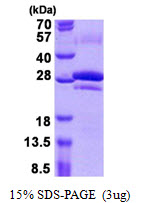
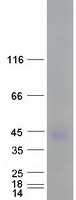
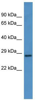
WB Suggested Anti-CEACAM7 Antibody Titration: 0.2-1 ug/ml; ELISA Titer: 1:62500; Positive Control: Jurkat cell lysate
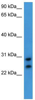
WB Suggested Anti-CEACAM7 Antibody Titration: 0.2-1 ug/ml; ELISA Titer: 1:62500; Positive Control: Hela cell lysate
- Quick-Filters:
- Cited (1)
Primary Antibodies
| Catalog No. | Host | Iso. | Clone | Pres. | React. | Applications | |
|---|---|---|---|---|---|---|---|
| DM1207 | CEACAM7 antibody |
||||||
 |
Mouse | IgG1 | BAC2 | Purified | Hu | C, E, F, ICC/IF |
0.1 mg /
€380.00
|
| OriGene Technologies GmbH | |||||||
 +1 additional image +1 additional image |
Published recently! |
||||||
Proteins & Growth Factors
| Catalog No. | Species | Pres. | Purity | Source | |
|---|---|---|---|---|---|
| TP312214 | CEACAM7 |
||||
| Purified recombinant protein of Homo sapiens carcinoembryonic antigen-related cell adhesion molecule 7 (CEACAM7) | |||||
 |
Human | > 80 % Preparation: Recombint protein was captured through anti-DDK affinity column followed by conventiol chromatography steps. Purity Detail: > 80% as determined by SDS-PAGE and Coomassie blue staining. |
HEK293 cells |
20 µg /
€748.00
|
|
| OriGene Technologies, Inc. | |||||