Complex I subunit NDUFS3 antibody
Principal name
Complex I subunit NDUFS3 antibody
Alternative names for Complex I subunit NDUFS3 antibody
Mitochondria Complex I (NADH Dehydrogenase), NADH-ubiquinone oxidoreductase 30 kDa subunit, Complex I-30kD, NADH dehydrogenase [ubiquinone] iron-sulfur protein 3, mitochondrial
Ncbi ID
9606, NP_004542, NP_004542.1, NM_004551, NP_080964.1, NM_026688
Available reactivities
Available hosts
Available applications
Enzyme Immunoassay (E), Western blot / Immunoblot (WB), Paraffin Sections (P), ELISA (detection) (E(detection)), Immunoprecipitation (IP)
Background of Complex I subunit NDUFS3 antibody
- Kit Component:
- - KN310856G1, Ndufs3 gRNA vector 1 in pCas-Guide vector
- KN310856G2, Ndufs3 gRNA vector 2 in pCas-Guide vector
- KN310856D, donor vector containing left and right homologous arms and Luciferase-Puro functional cassette
- GE100003, scramble sequence in pCas-Guide vector - CRISPR Manual:
- CRISPR/CAS9 Genome Editing Manual
General readings
Murray J., et al. (2003) J. Biol. Chem. 278:13619-13622Saada A., et al. (2009) Am. J. Hum. Genet. 84:718-727
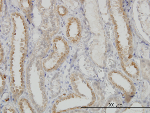
Immunoperoxidase of monoclonal antibody to NDUFS3 on formalin-fixed paraffin-embedded human kidney. [antibody concentration 3 ug/ml]

Western blot analysis of NDUFS3 over-expressed 293 cell line, cotransfected with NDUFS3 Validated Chimera RNAi ( Cat # H00004722-R01V ) (Lane 2) or non-transfected control (Lane 1). Blot probed with NDUFS3 monoclonal antibody (M02), clone 1D6 (Cat # H00004722-M02 ). GAPDH ( 36.1 kDa ) used as specificity and loading control.
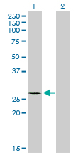
Western Blot analysis of NDUFS3 expression in transfected 293T cell line by NDUFS3 monoclonal antibody (M02), clone 1D6.Lane 1: NDUFS3 transfected lysate(30.2 KDa).Lane 2: Non-transfected lysate.

NDUFS3 MaxPab polyclonal antibody. Western Blot analysis of NDUFS3 expression in rat brain.
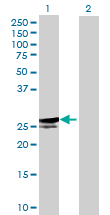
Western Blot analysis of NDUFS3 expression in transfected 293T cell line (H00004722-T01) by NDUFS3 MaxPab polyclonal antibody.Lane 1: NDUFS3 transfected lysate(29.04 KDa).Lane 2: Non-transfected lysate.
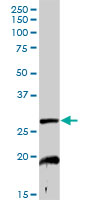
NDUFS3 MaxPab rabbit polyclonal antibody. Western Blot analysis of NDUFS3 expression in mouse kidney.
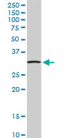
NDUFS3 MaxPab rabbit polyclonal antibody. Western Blot analysis of NDUFS3 expression in human pancreas.

Western Blot analysis of NDUFS3 expression in transfected 293T cell line (H00004722-T02) by NDUFS3 MaxPab polyclonal antibody.Lane 1: NDUFS3 transfected lysate(30.20 KDa).Lane 2: Non-transfected lysate.
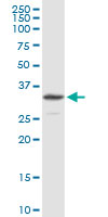
NDUFS3 MaxPab rabbit polyclonal antibody. Western Blot analysis of NDUFS3 expression in HepG2.
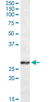
NDUFS3 polyclonal antibody ( Cat # PAB14140 ) ( 0.3µg/ml ) staining of human frontal cortex lysate ( 35µg protein in RIPA buffer ). Primary incubation was 1 hour. Detected by chemiluminescence.
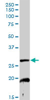
NDUFS3 MaxPab rabbit polyclonal antibody. Western Blot analysis of NDUFS3 expression in mouse kidney.

NDUFS3 MaxPab rabbit polyclonal antibody. Western Blot analysis of NDUFS3 expression in HepG2.
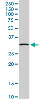
NDUFS3 MaxPab rabbit polyclonal antibody. Western Blot analysis of NDUFS3 expression in human pancreas.

Western Blot analysis of NDUFS3 expression in transfected 293T cell line (H00004722-T02) by NDUFS3 MaxPab polyclonal antibody.Lane 1: NDUFS3 transfected lysate(30.20 KDa).Lane 2: Non-transfected lysate.
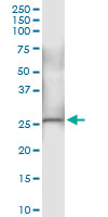
Immunoprecipitation of NDUFS3 transfected lysate using anti-NDUFS3 MaxPab rabbit polyclonal antibody and Protein A Magnetic Bead (U0007), and immunoblotted with NDUFS3 MaxPab mouse polyclonal antibody (B01) (H00004722-B01).

Human PANC1; WB Suggested Anti-NDUFS3 Antibody Titration: 0.2-1 ug/ml. Positive Control: PANC1 cell lysate; NDUFS3 antibody - middle region (ARP56578_P050) in Human PANC1 cells using Western Blot
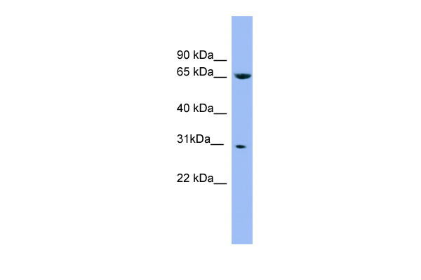
Human Placenta; WB Suggested Anti-NDUFS3 Antibody Titration: 0.2-1 ug/ml. ELISA Titer: 1:1562500. Positive Control: Human Placenta; NDUFS3 antibody - middle region (ARP56579_P050) in Human Placenta cells using Western Blot
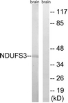

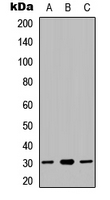
Western blot analysis of NDUFS3 expression in HeLa (A); HepG2 (B); HEK293T (C) whole cell lysates.
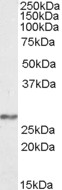
TA305654 (0.3ug/ml) staining of Human Frontal Cortex lysate (35ug protein in RIPA buffer). Primary incubation was 1 hour. Detected by chemiluminescence.
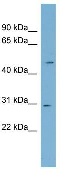
WB Suggested Anti-NDUFS3 Antibody Titration: 0.2-1 ug/ml; Positive Control: PANC1 cell lysate

WB Suggested Anti-NDUFS3 Antibody Titration: 0.2-1 ug/ml; ELISA Titer: 1:1562500; Positive Control: Human Placenta
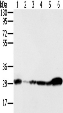
Gel: 8%SDS-PAGE, Lysate: 40 ug, Lane 1-6: 293T cells, NIH/3T3 cells, Raw264.7 cells, hela cells, human fetal brain tissue, mouse brain tissue, Primary antibody: (NDUFS3 Antibody) at dilution 1/350, Secondary antibody: Goat anti rabbit IgG at 1/8000 dilution, Exposure time: 5 seconds
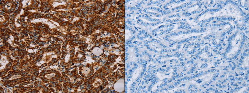
The image on the left is immunohistochemistry of paraffin-embedded Human thyroid cancer tissue using (NDUFS3 Antibody) at dilution 1/40, on the right is treated with fusion protein. (Original magnification: ×200)
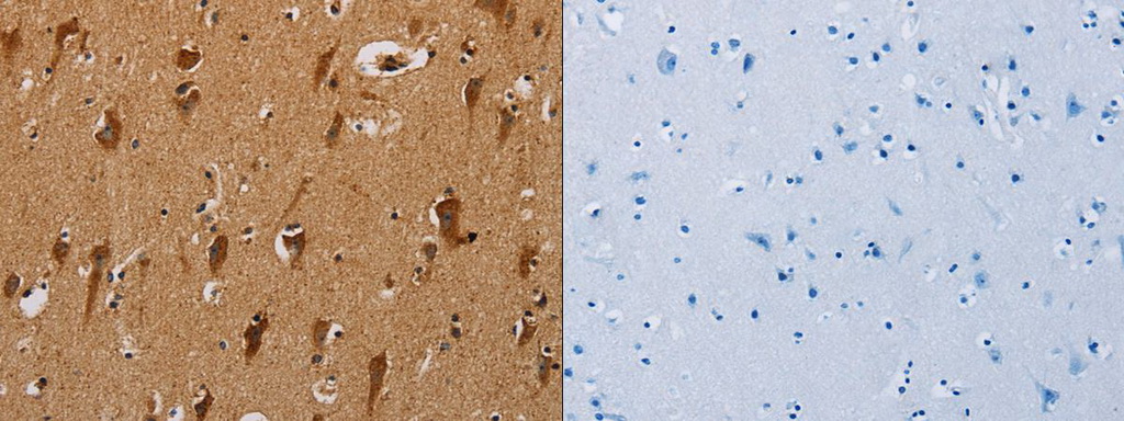
The image on the left is immunohistochemistry of paraffin-embedded Human brain tissue using (NDUFS3 Antibody) at dilution 1/40, on the right is treated with fusion protein. (Original magnification: ×200)
Primary Antibodies
| Catalog No. | Host | Iso. | Clone | Pres. | React. | Applications | |
|---|---|---|---|---|---|---|---|
| TA305654 | Complex I subunit NDUFS3 antibody |
||||||
| Goat Anti-NDUFS3 Antibody | |||||||
 |
Goat | Aff - Purified | Bov, Hu | WB |
0.1 mg /
€360.00
|
||
| OriGene Technologies, Inc. | |||||||