Ephrin-A3 antibody
Principal name
Ephrin-A3 antibody
Alternative names for Ephrin-A3 antibody
EFNA3, LERK-3, EHK1 ligand, EHK1-L, EFL-2, EFL2, EPLG3, LERK3, EPH-related receptor tyrosine kinase ligand 3
Ncbi ID
Available reactivities
Available hosts
Available applications
Enzyme Immunoassay (E), Western blot / Immunoblot (WB), ELISA (detection) (E(detection)), Immunocytochemistry/Immunofluorescence (ICC/IF), Paraffin Sections (P), Flow Cytometry (F)
Background of Ephrin-A3 antibody
- Kit Component:
- - KN210313G1, EFNA3 gRNA vector 1 in pCas-Guide vector
- KN210313G2, EFNA3 gRNA vector 2 in pCas-Guide vector
- KN210313D, donor vector containing left and right homologous arms and Luciferase-Puro functional cassette
- GE100003, scramble sequence in pCas-Guide vector - CRISPR Manual:
- CRISPR/CAS9 Genome Editing Manual
General readings
Carmona MA, Murai KK, et al. Proc Natl Acad Sci U S A. 2009 Jul 28;106(30):12524-9.
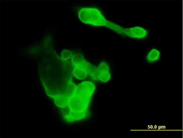
Immunofluorescence of purified MaxPab antibody to EFNA3 on 293 cell. [antibody concentration 1 ug/ml]
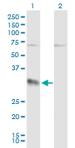
Western Blot analysis of EFNA3 expression in transfected 293T cell line (H00001944-T02) by EFNA3 MaxPab polyclonal antibody.Lane 1: EFNA3 transfected lysate(26.40 KDa).Lane 2: Non-transfected lysate.
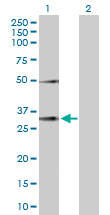
Western Blot analysis of EFNA3 expression in transfected 293T cell line (H00001944-T01) by EFNA3 MaxPab polyclonal antibody.Lane 1: EFNA3 transfected lysate(26.40 KDa).Lane 2: Non-transfected lysate.
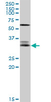
EFNA3 MaxPab rabbit polyclonal antibody. Western Blot analysis of EFNA3 expression in Jurkat.
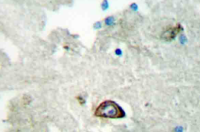
Immunohistochemistry (IHC) analyzes of Ephrin-A3 antibody (Cat.-No.: AP20385PU-N) in paraffin-embedded human brain tissue.
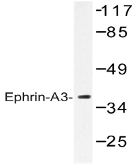
Western blot (WB) analysis of Ephrin-A3 antibody (Cat.-No.: AP20385PU-N) in extracts from HepG2 cells.

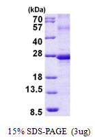
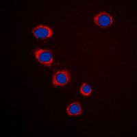
Immunofluorescent analysis of Ephrin A3 staining in HepG2 cells. Formalin-fixed cells were permeabilized with 0.1% Triton X-100 in TBS for 5-10 minutes and blocked with 3% BSA-PBS for 30 minutes at room temperature. Cells were probed with the primary antibody in 3% BSA-PBS and incubated overnight at 4 C in a humidified chamber. Cells were washed with PBST and incubated with a DyLight 594-conjugated secondary antibody (red) in PBS at room temperature in the dark. DAPI was used to stain the cell nuclei (blue).
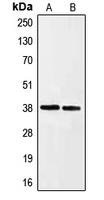
Western blot analysis of Ephrin A3 expression in HeLa (A); HepG2 (B) whole cell lysates.
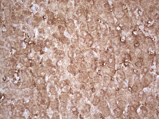
Immunohistochemical staining of paraffin-embedded Human liver tissue within the normal limits using anti-EFNA3 mouse monoclonal antibody. (Heat-induced epitope retrieval by 1 mM EDTA in 10mM Tris, pH8.5, 120C for 3min, TA806050)
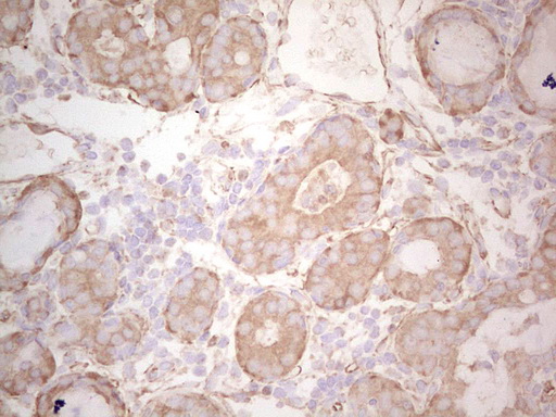
Immunohistochemical staining of paraffin-embedded Adenocarcinoma of Human breast tissue using anti-EFNA3 mouse monoclonal antibody. (Heat-induced epitope retrieval by 1 mM EDTA in 10mM Tris, pH8.5, 120C for 3min, TA806050)
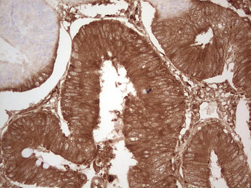
Immunohistochemical staining of paraffin-embedded Adenocarcinoma of Human colon tissue using anti-EFNA3 mouse monoclonal antibody. (Heat-induced epitope retrieval by 1 mM EDTA in 10mM Tris, pH8.5, 120C for 3min, TA806050)
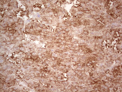
Immunohistochemical staining of paraffin-embedded Carcinoma of Human liver tissue using anti-EFNA3 mouse monoclonal antibody. (Heat-induced epitope retrieval by 1 mM EDTA in 10mM Tris, pH8.5, 120C for 3min, TA806050)
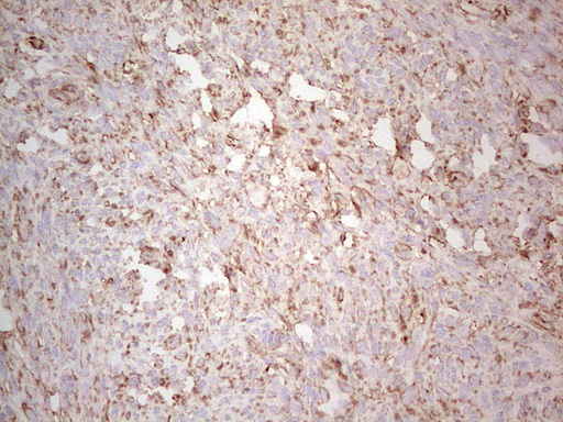
Immunohistochemical staining of paraffin-embedded Human endometrium tissue within the normal limits using anti-EFNA3 mouse monoclonal antibody. (Heat-induced epitope retrieval by 1 mM EDTA in 10mM Tris, pH8.5, 120C for 3min, TA806050)
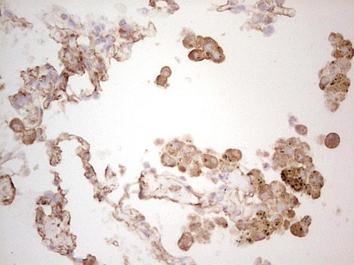
Immunohistochemical staining of paraffin-embedded Human lung tissue within the normal limits using anti-EFNA3 mouse monoclonal antibody. (Heat-induced epitope retrieval by 1 mM EDTA in 10mM Tris, pH8.5, 120C for 3min, TA806050)
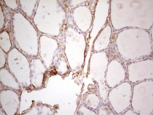
Immunohistochemical staining of paraffin-embedded Human thyroid tissue within the normal limits using anti-EFNA3 mouse monoclonal antibody. (Heat-induced epitope retrieval by 1 mM EDTA in 10mM Tris, pH8.5, 120C for 3min, TA806050)
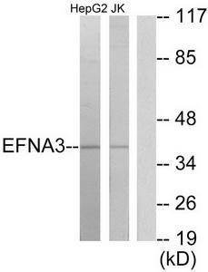
Western blot analysis of extracts from HepG2 cells and Jurkat cells, using EFNA3 antibody.The lane on the right is treated with the synthesized peptide.
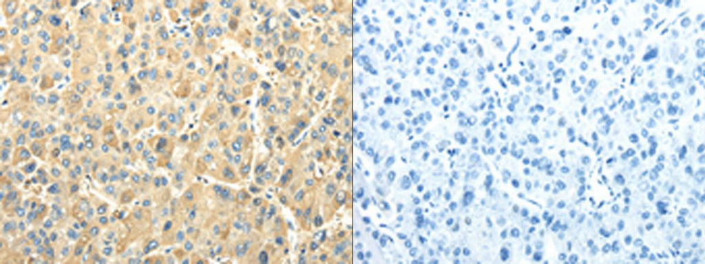
The image on the left is immunohistochemistry of paraffin-embedded Human liver cancer tissue using EFNA3 Antibody at dilution 1/25, on the right is treated with fusion protein. (Original magnification: ×200)
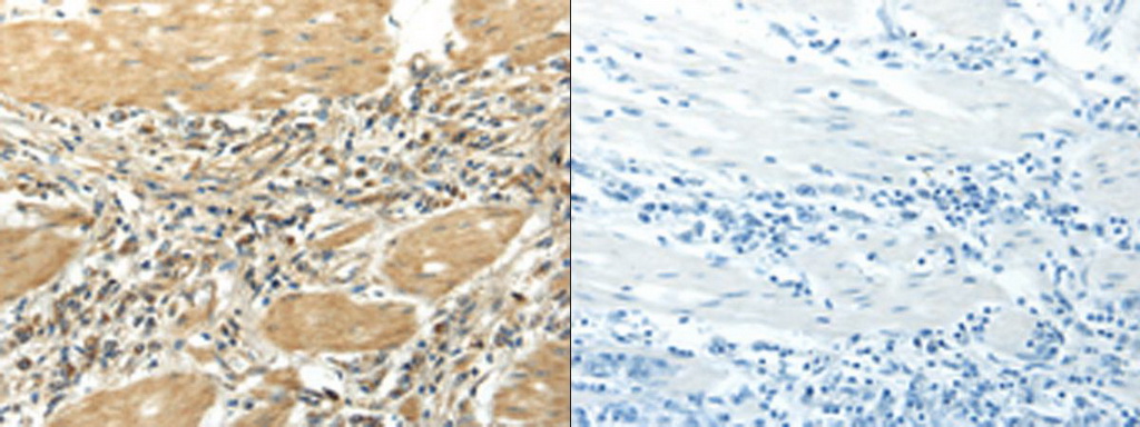
The image on the left is immunohistochemistry of paraffin-embedded Human gastric cancer tissue using EFNA3 Antibody at dilution 1/25, on the right is treated with fusion protein. (Original magnification: ×200)
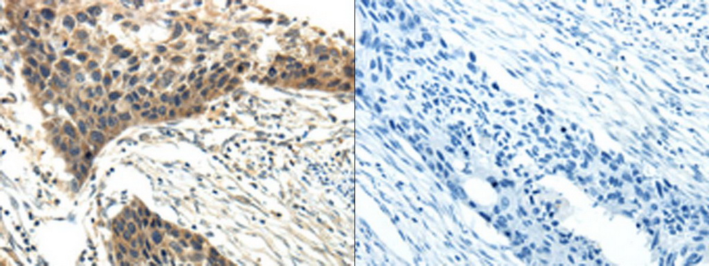
The image on the left is immunohistochemistry of paraffin-embedded Human esophagus cancer tissue using EFNA3 Antibody at dilution 1/20, on the right is treated with fusion protein. (Original magnification: ×200)
- Quick-Filters:
- Highly Validated (1)
Primary Antibodies
| Catalog No. | Host | Iso. | Clone | Pres. | React. | Applications | |
|---|---|---|---|---|---|---|---|
| TA806050 | Ephrin-A3 antibody |
||||||
| EFNA3 mouse monoclonal antibody, clone OTI4A4 (formerly 4A4) | |||||||
 |
Mouse | IgG2b | OTI4A4 | Purified | Hu | P |
0.1 ml /
€360.00
|
| OriGene Technologies, Inc. | |||||||
 +6 additional images +6 additional images |
Highly validated antibody!
|
||||||
| TA311683 | Ephrin-A3 antibody |
||||||
| Rabbit polyclonal anti-EFNA3 antibody | |||||||
 |
Rabbit | IgG | Purified | Hu, Ms, Rt | E, WB |
0.1 ml /
€335.00
|
|
| OriGene Technologies, Inc. | |||||||
| TA322696 | Ephrin-A3 antibody |
||||||
| Anti-EFNA3 Rabbit Polyclonal Antibody | |||||||
 |
Rabbit | IgG | Purified | Hu, Ms, Rt | E, WB |
0.1 ml /
€335.00
|
|
| OriGene Technologies, Inc. | |||||||
| +1 additional image |
|||||||
| TA322697 | Ephrin-A3 antibody |
||||||
| Anti-EFNA3 Rabbit Polyclonal Antibody | |||||||
 |
Rabbit | IgG | Purified | Hu, Ms, Rt | E, WB |
0.1 ml /
€335.00
|
|
| OriGene Technologies, Inc. | |||||||
| AP20385PU-N | Ephrin-A3 antibody |
||||||
 |
Rabbit | Aff - Purified | Hu, Ms, Rt | ICC/IF, P, WB |
0.1 mg /
€370.00
|
||
| OriGene Technologies GmbH | |||||||
| +1 additional image |
|||||||