GFP antibody
Principal name
GFP antibody
Alternative names for GFP antibody
Green fluorescent protein, GFP-Tag
SwissProt ID
P42212 (Aeqvi)
Gene ID
3020 (H3F3A)
Available reactivities
Available hosts
Available applications
Western blot / Immunoblot (WB), Frozen Sections (C), Immunoprecipitation (IP), Immunocytochemistry/Immunofluorescence (ICC/IF), Enzyme Immunoassay (E), Paraffin Sections (P), ChIP assay (ChIP), In Situ Hybridization (ISH), Immunoelectron Microscopy (EM), Dot blot (Dot), Flow Cytometry (F), Immunohistochemistry (IHC)
Background of GFP antibody
Green fluorescence protein (GFP) is a 27 KDa protein derived from the bioluminiscent jellyfish Aquorea victoria, emiting green light (509 nm) when excited (excitation by Blue or UV light, absorption peak at 395 nm).
GFP is a useful tool in cell biology research, as its intrinsic fluorescence can be visualized in living cells. Light-stimulated GFP fluorescence is species-independent and a fluorescence has been reported from many different types of GFP-expressing hosts, including microbes, invertebrates, vertebrates and plants. No exogenous substrates and cofactors are required for the fluorescence of GFP, since GFP autocatalytically forms a fluorescent pigment from natural amino acids present in the nascent protein.
GFP fluorescence is stable under fixation conditions and suitable for a variety of applications. GFP is widely used as a reporter (tag) for gene expression, enabling researchers to visualize and localize GFP-tagged proteins within living cells without any further staining. Other applications of GFP include measurement of distance between proteins through fluorescence energy transfer (FRET) protocols.
To increase a fluorescence intensity of GFP, chromophore mutations have been created. The Enhanced GFP has a fluorescence 35 times more intense than the wt-GFP. Mutagenesis of GFP has produced also many mutants (e.g. Yellow Fluorescent Protein, Cyan Fluorescent Protein) with warying spectral properties. Antibodies raised against full-length GFP variants should also detect other variants of the protein.
General readings
Schoch KG et al. A subset of mouse tracheal epithelial basal cells generates large colonies in vitro. Am J Physiol Lung Cell Mol Physiol 286:L631-42 (2004). PubMed PMID: 12959927
Karabay A. et al Axonal growth is sensitive to the levels of katanin, a protein that severs microtubules. The Journal of Neuroscience 24:5778-5788 (2004).Gongidi V et al. SPARC-like 1 Regulates the Terminal Phase of Radial Glia-Guided Migration in the Cerebral Cortex. Neuron 41:57-69 (2004). PubMed PMID: 14715135
Fischer AC et al. Successful transgene expression with serial doses of aerosolized rAAV2 vectors in rhesus macaques. Mol Ther 8:918-26 (2003). PubMed PMID: 14664794
Wong JM et al. Subnuclear shuttling of human telomerase induced by transformation and DNA damage. Nat Cell Biol 4:731-6 (2002). PubMed PMID: 12198499
Zhang QX et al. Identification of structurally important domains of lipid phosphate phosphatase-1: implications for its sites of action. Biochem J 345 Pt 2:181-4 (2000). PubMed PMID: 10620492
Mesaeli N et al. Calreticulin is essential for cardiac development. J Cell Biol 144:857-68 (1999). PubMed PMID: 10085286
McCabe JB & Berthiaume LG Functional roles for fatty acylated amino-terminal domains in subcellular localization. Mol Biol Cell 10:3771-86 (1999). PubMed PMID: 10564270
Jasinska R et al. Lipid phosphate phosphohydrolase-1 degrades exogenous glycerolipid and sphingolipid phosphate esters. Biochem J 340 ( Pt 3):677-86 (1999). PubMed PMID: 10359651
1. Reinhardt, R. L., Hong, S., Kang, S.-J., Wang, Z.-e., and Locksley, R. M. (2006) Visualization of IL-12/23p40 In Vivo Reveals Immunostimulatory Dendritic Cell Migrants that Promote Th1 Differentiation, 177, 1618-1627.
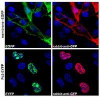
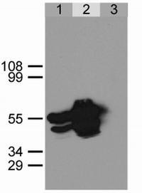
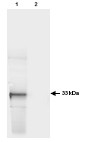

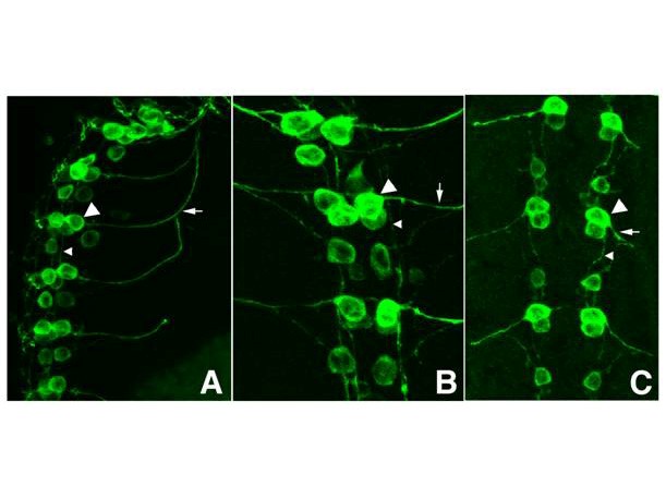
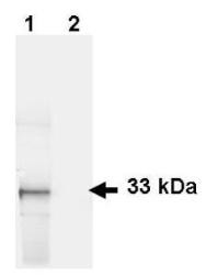
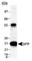
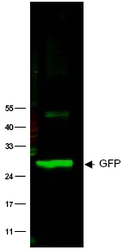

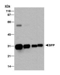
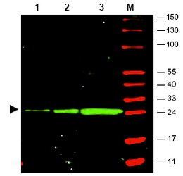



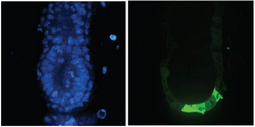
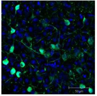

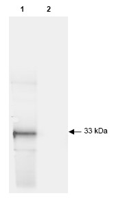
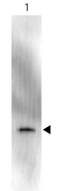
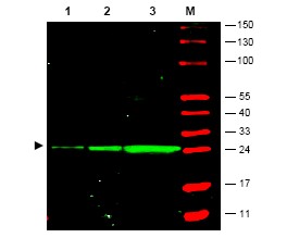

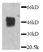
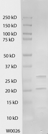
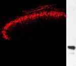
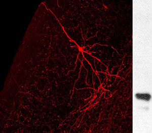

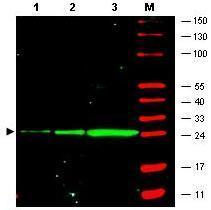
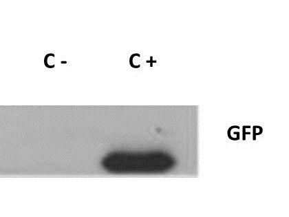
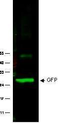

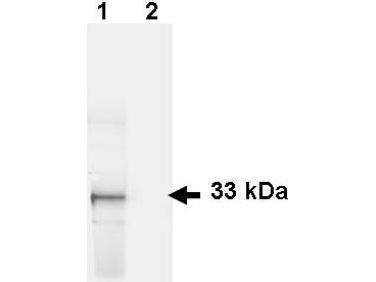

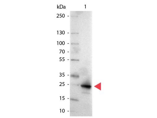
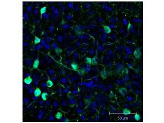

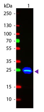
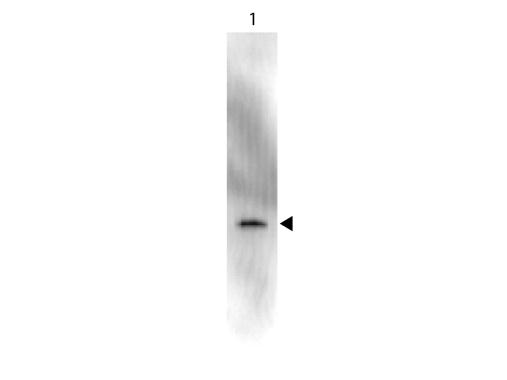

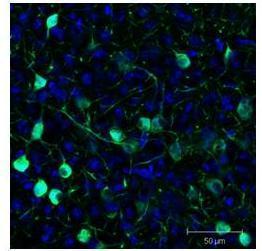
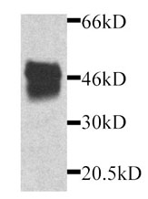
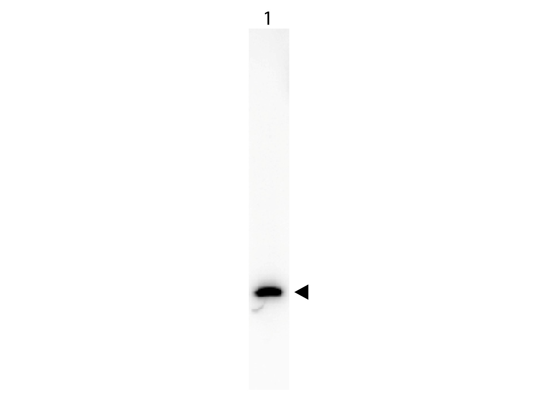
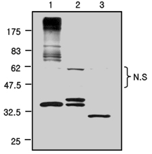
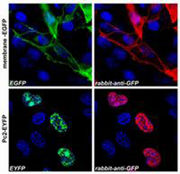
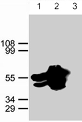
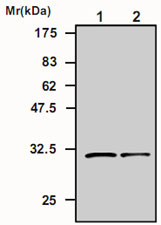
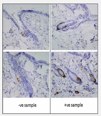

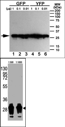
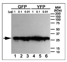
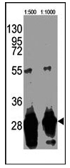

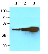
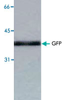
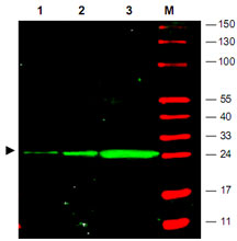
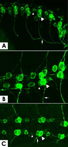
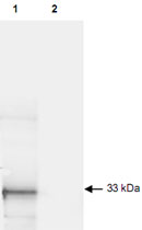
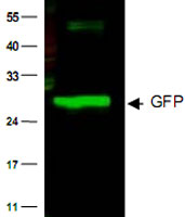
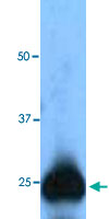

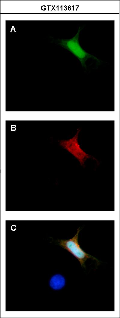

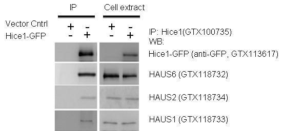
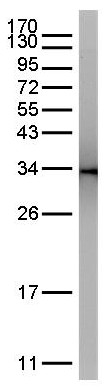


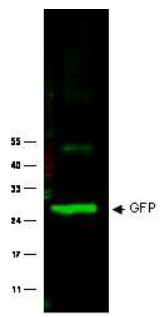
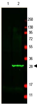
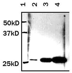
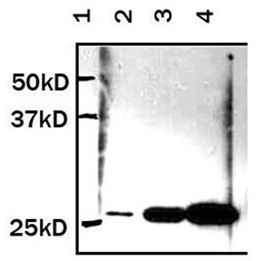
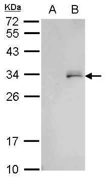
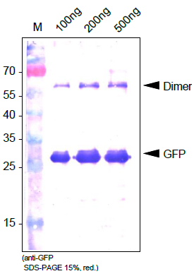
Predicted/Observed size: 27 kDa for epitope tag GFP. Other band(s): none.
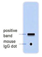
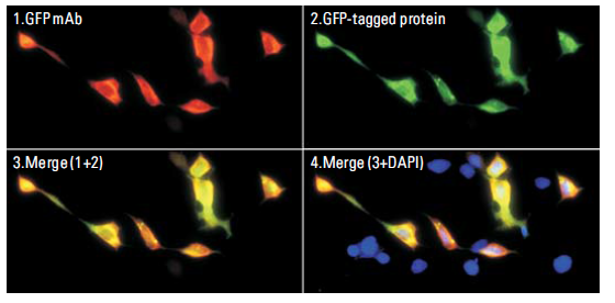
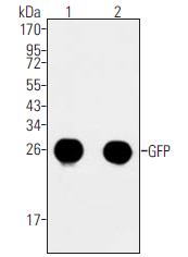
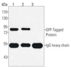
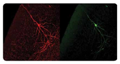


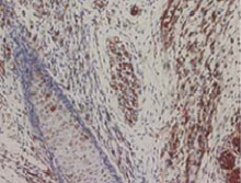
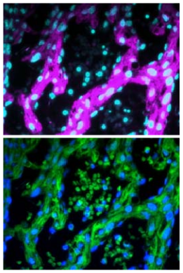
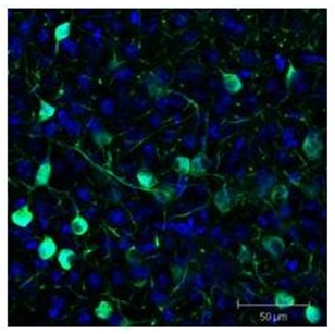
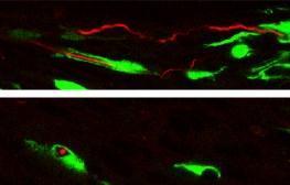
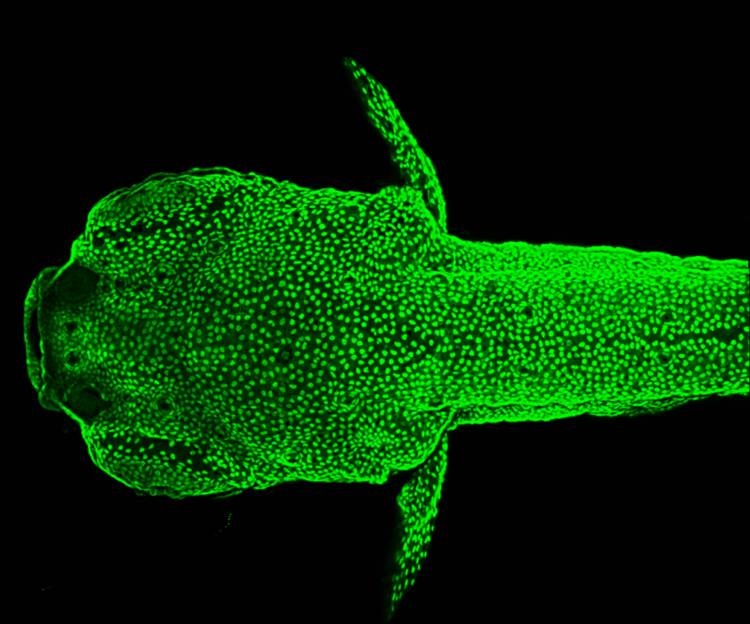
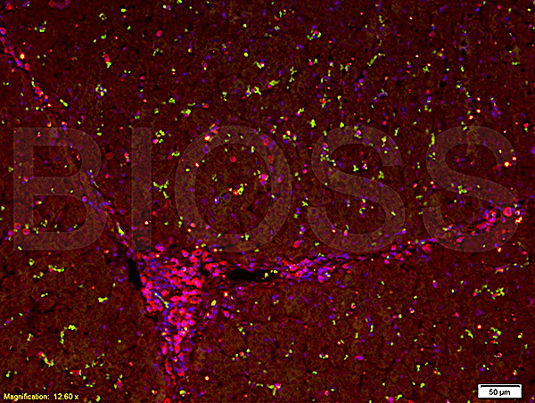
Magenta: AM26599AF-N.
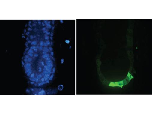
Green: GFP own fluorescence.
Cyan, Blue: DAP.
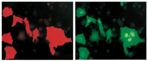
Immunocytochemical detection of GFP on 4% PFA fixed transfectant using AM26599AF-N (left). Right panel is GFP own fluorescence.
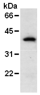
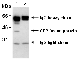
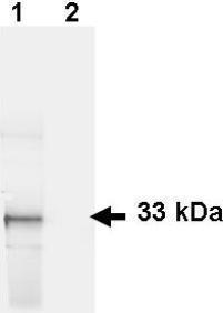
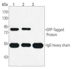
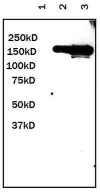
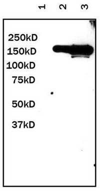
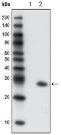
Lane 1: IP with AM26599AF-N.
Lane 2: IP with isotype control.
- Quick-Filters:
- Cited (14)
- Highly Validated (3)
- Overnight Delivery (6)

Primary Antibodies
| Catalog No. | Host | Iso. | Clone | Pres. | React. | Applications | |
|---|---|---|---|---|---|---|---|
| SP3005P | GFP antibody |
||||||
 |
Rabbit | Purified | All Species | ICC/IF, IP, WB |
0.1 mg /
€230.00
|
||
| OriGene Technologies GmbH | |||||||
 +1 additional image +1 additional image |
Top Selling product! Published recently! |
||||||
| AM11009PU-N | GFP antibody |
||||||
 |
Mouse | IgG1 | 168AT1211 | Purified | ICC/IF, P, WB |
0.4 ml /
€415.00
|
|
| OriGene Technologies GmbH | |||||||
| +2 additional images |
Published recently! |
||||||
| AM11010SU-N | GFP antibody |
||||||
 |
Mouse | IgG | 168AT1211.269.64 | Ascites | WB |
0.1 ml /
€415.00
|
|
| OriGene Technologies GmbH | |||||||
| +1 additional image |
Published recently! |
||||||
| AP09218PU-N | GFP (Ads. to Hu, Ms, Rt Serum Proteins) antibody |
||||||
![Western blot of GFP protein detected with polyclonal anti-GFP antibody. Wild type GFP (0.1 μg) was used to spike 30 μg of a HeLa whole cell lysate. This antibody detects a 27 kDa band corresponding to the epitope tag GFP. A 4-20% Tris-Glycine gradient gel was used for SDS-PAGE. The protein was transferred to nitrocellulose using standard methods. After blocking with 5% BLOTTO in PBS, the membrane was probed overnight at 4° C with the primary antibody diluted in 5% BLOTTO to 1:1,000, followed by washes and reaction with a 1:10,000 dilution of IRDye® 800 conjugated Goat-a-Rabbit IgG [H&L]. IRDye® 800 fluorescence image was captured using the Odyssey® Infrared Imaging System developed by LI-COR. IRDye is a trademark of LI-COR, Inc. Other detection systems will yield similar results.](https://m1.acris-antibodies.com/catalog/product/cache/1/small_image/50x50/9df78eab33525d08d6e5fb8d27136e95/0163839.png) |
Rabbit | IgG | Aff - Purified | A. victoria | C, E, ICC/IF, WB |
0.1 mg /
€390.00
|
|
| OriGene Technologies GmbH | |||||||
| +2 additional images |
Published recently! |
||||||
| AP54983SU-N | GFP antibody |
||||||
 |
Rabbit | Serum | C, E, ICC/IF, WB |
0.2 ml /
€380.00
|
|||
| OriGene Technologies GmbH | |||||||
Published recently! |
|||||||
| R1091B | GFP (Ads. to Hu, Ms, Rt Serum Proteins) antibody |
||||||
 |
Goat | Biotin | A. victoria | C, E, ICC/IF, WB |
1 mg /
€630.00
|
||
| OriGene Technologies GmbH | |||||||
| +2 additional images |
Published recently! |
||||||
| R1091F | GFP antibody |
||||||
 |
Goat | FITC | A. victoria | C, E, ICC/IF, WB |
1 mg /
€640.00
|
||
| OriGene Technologies GmbH | |||||||
Published recently! |
|||||||
| R1091P | GFP (Ads. to Hu, Ms, Rt Serum Proteins) antibody |
||||||
 |
Goat | Aff - Purified | C, E, ICC/IF, WB |
1 mg /
€600.00
|
|||
| OriGene Technologies GmbH | |||||||
| +3 additional images |
Published recently! Highly validated antibody! |
||||||
| R1091T | GFP antibody |
||||||
|
Goat | TRITC | E, ICC/IF, WB |
1 mg /
€610.00
|
|||
| OriGene Technologies GmbH | |||||||
Published recently! |
|||||||
| R1091TR | GFP antibody |
||||||
|
Goat | Texas Red | C, ICC/IF |
1 mg /
€630.00
|
|||
| OriGene Technologies GmbH | |||||||
Published recently! |
|||||||
| R1461HRP | GFP antibody |
||||||
 |
Mouse | IgG2a | 9F9.F9 | HRP | A. victoria | C, E, WB |
1 mg /
€855.00
|
| OriGene Technologies GmbH | |||||||
Published recently! |
|||||||
| AM06180PU-N | GFP antibody |
||||||
 |
Mouse | IgG2a | 4B10B2 | Purified | E, F, P, WB |
0.1 ml /
€340.00
|
|
| OriGene Technologies GmbH | |||||||
| +3 additional images |
Highly validated antibody! |
||||||
| AM26599AF-N | GFP antibody |
||||||
 |
Mouse | IgG2b | 1E4 | Azide Free | ICC/IF, IP, P, WB |
0.1 mg /
€355.00
|
|
| OriGene Technologies GmbH | |||||||
| +4 additional images |
Highly validated antibody! |
||||||
| 73-131 | GFP antibody |
||||||
 |
Mouse | IgG2a | N86/8 | Supernatant | C, ICC/IF, IP, WB |
5 ml /
€490.00
|
|
| Antibodies Incorporated | |||||||
| +2 additional images |
|
||||||
| 73-132 | GFP antibody |
||||||
 |
Mouse | IgG2a | N86/38 | Supernatant | C, ICC/IF, IP, WB |
5 ml /
€490.00
|
|
| Antibodies Incorporated | |||||||
| +2 additional images |
|
||||||
| 75-131 | GFP antibody |
||||||
 |
Mouse | IgG2a | N86/8 | Purified | C, ICC/IF, IP, WB |
0.1 ml /
€570.00
|
|
| Antibodies Incorporated | |||||||
| +2 additional images |
|
||||||
| 75-132 | GFP antibody |
||||||
 |
Mouse | IgG2a | N86/38 | Purified | C, ICC/IF, IP, WB |
0.1 ml /
€570.00
|
|
| Antibodies Incorporated | |||||||
| +2 additional images |
|
||||||
| AB0020-20 | GFP antibody |
||||||
 |
Goat | IgG | Aff - Purified | C, ICC/IF, P, WB |
20 µl /
€50.00
|
||
| SICGEN | |||||||
| AB0020-200 | GFP antibody |
||||||
|
Goat | IgG | Aff - Purified | C, ICC/IF, P, WB |
0.4 mg /
€210.00
|
||
| SICGEN | |||||||
| AB0020-500 | GFP antibody |
||||||
|
Goat | IgG | Aff - Purified | C, ICC/IF, P, WB |
1.0 mg /
€420.00
|
||
| SICGEN | |||||||

