HAUS8 antibody
Principal name
HAUS8 antibody
Alternative names for HAUS8 antibody
HICE1, Sarcoma antigen NY-SAR-48, HAUS augmin-like complex subunit 8, HEC1/NDC80-interacting centrosome-associated protein 1
SwissProt ID
Q9BT25 (Human)
Gene ID
93323 (HAUS8)
Ncbi ID
Available reactivities
Available hosts
Available applications
ELISA (detection) (E(detection)), Western blot / Immunoblot (WB), Immunocytochemistry/Immunofluorescence (ICC/IF), Immunoprecipitation (IP), Paraffin Sections (P), Enzyme Immunoassay (E)
Background of HAUS8 antibody
- Kit Component:
- - KN217063G1, HAUS8 gRNA vector 1 in pCas-Guide vector
- KN217063G2, HAUS8 gRNA vector 2 in pCas-Guide vector
- KN217063D, donor vector containing left and right homologous arms and Luciferase-Puro functional cassette
- GE100003, scramble sequence in pCas-Guide vector - CRISPR Manual:
- CRISPR/CAS9 Genome Editing Manual
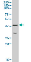
NY-SAR-48 MaxPab polyclonal antibody. Western Blot analysis of NY-SAR-48 expression in human spleen.
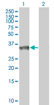
Western Blot analysis of HICE1 expression in transfected 293T cell line (H00093323-T01) by HICE1 MaxPab polyclonal antibody.Lane 1: NY-SAR-48 transfected lysate(27.39 KDa).Lane 2: Non-transfected lysate.
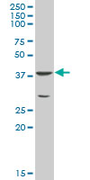
NY-SAR-48 MaxPab polyclonal antibody. Western Blot analysis of NY-SAR-48 expression in human spleen.
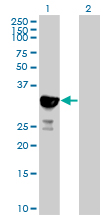
Western Blot analysis of HICE1 expression in transfected 293T cell line (H00093323-T01) by HICE1 MaxPab polyclonal antibody.Lane 1: NY-SAR-48 transfected lysate(27.39 KDa).Lane 2: Non-transfected lysate.

Human HeLa; WB Suggested Anti-HAUS8 Antibody Titration: 0.125ug/ml. ELISA Titer: 1:312500. Positive Control: Hela cell lysate; HAUS8 antibody - N-terminal region (ARP59892_P050) in Human HeLa cells using Western Blot
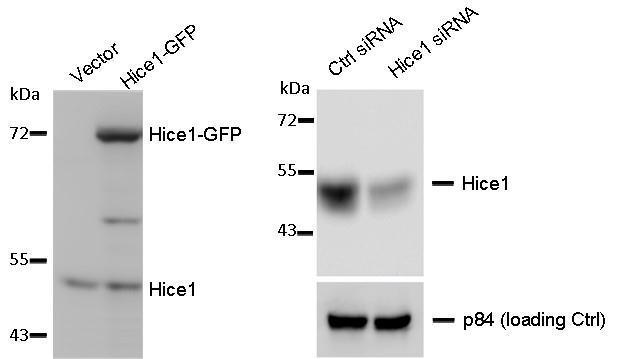
WB to detect cellular Hice1 and Hice1-GFP expressed in human osteosarcoma U2OS cells (left image), and Hice1 upon siRNA treatment (right image), using TA307936 at 1:1000 dilution. Nuclear matrix protein p84 is a loading control, blotted with p84 antibody
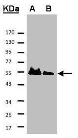
Sample. A: His-Hice1 (2x). B: His-Hice1 (1x). 7.5% SDS PAGE. TA307936 diluted at 1:500
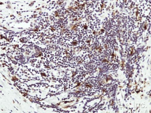
Immunohistochemical analysis of paraffin-embedded Human lymph tissue, using HICE1 (TA307936) antibody at 1:50 dilution.
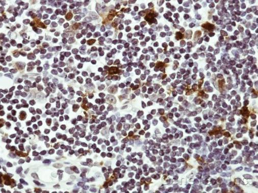
Immunohistochemical analysis of paraffin-embedded Human lymph tissue, using HICE1 (TA307936) antibody at 1:50 dilution.
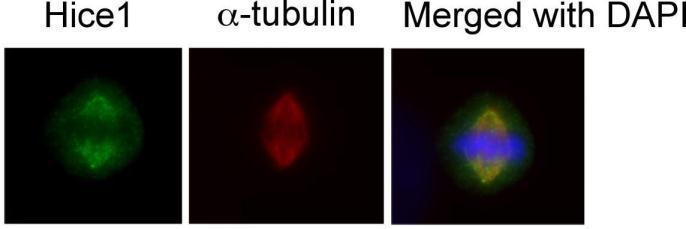
Immunofluorescence analysis of human osteosarcoma cell line U2OS, using HICE1(TA307936) antibody at 1:50-1:200 dilution.
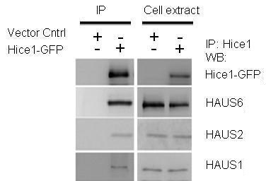
IP-WB assay to show that Hice1 co-immunoprecipitated with other Augmin components HAUS6, HAUS2 and HAUS1 in U2OS cells.

WB of Rabbit Anti-Hice1 pS70 antibody. Lane 1: HeLa cell extracts of untransfected cells (-). Lane 2: transfected HeLa cell extracts with Flag X3-Hice1 WT (WT). Lane 3: transfected HeLa cell extracts with Flag X3-Hice1 S70A mutant (70A). Load: 35 µg per lane. Primary antibody: Hice1 pS70 antibody at 0.5 µg/mL for overnight at 4°C. Secondary antibody: IRDye800™ Conjugated Goat Anti-Rabbit IgG secondary antibody at 1:10,000 for 45 min at RT.
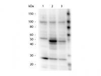
Western Blot of Rabbit anti-HICE1 pS70 antibody. Lane 1: 293T Null. Lane 2: 293T WT Hice1. Lane 3: 293T S70A Hice1. Load: 14 µl per lane. Primary antibody: HICE1 pS70 antibody at 0.75 ug/mL overnight at 4°C. Secondary antibody: Peroxidase rabbit secondary antibody at 1:40,000 for 30 min at RT. Block: MB-070 for 30 min at RT. Predicted/Observed size: 48 kDa, 48 kDa for HICE1 pS70. Other band(s): HICE1 pS70 splice variants and isoforms.

Anti-HICE1 in WB using Immunochemicals' Anti-HICE1 Antibody shows detection of a 45 kDa band corresponding to endogenous HICE1 in lysates of S phase HeLa cells silenced for either control Luciferase or HICE1. In right lane (HICE1i): lysates from sh-HICE1 RNAi-treated lentivirus-infected cells. In left lane (GLi): lysates from sh-Luciferase lentivirus-infected cells as control. Anti-HICE1 Antibody was used at 1:10,000. Molecular weight estimation was made by comparison by prestained MW markers.

WB of Rabbit Anti-Hice1 antibody. Lane 1: HeLa cell extracts of untransfected cells (-). Lane 2: transfected HeLa cell extracts with Flag X3-Hice1 WT (WT). Lane 3: transfected HeLa cell extracts with Flag X3-Hice1 S70A mutant (70A). Load: 35 µg per lane. Primary antibody: Hice1 antibody at 0.5 µg/mL for overnight at 4°C. Secondary antibody: IRDye800™ Conjugated Goat Anti-Rabbit IgG secondary antibody at 1:10,000 for 45 min at RT.
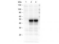
Western Blot of Rabbit anti-HICE1 antibody. Lane 1: 293T Null. Lane 2: 293T WT Hice1. Lane 3: 293T S70A Hice1. Load: 14 µl per lane. Primary antibody: HICE1 antibody at 1.24 ug/mL overnight at 4°C. Secondary antibody: Peroxidase rabbit secondary antibody at 1:40,000 for 30 min at RT. Block: MB-070 for 30 min at RT. Predicted/Observed size: 48 kDa, 48 kDa for HICE1. Other band(s): HICE1 splice variants and isoforms.
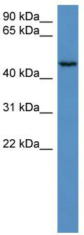
WB Suggested Anti-HAUS8 Antibody Titration: 0.125 ug/ml; ELISA Titer: 1:312500; Positive Control: Hela cell lysate
- Quick-Filters:
- Highly Validated (1)
Primary Antibodies
| Catalog No. | Host | Iso. | Clone | Pres. | React. | Applications | |
|---|---|---|---|---|---|---|---|
| TA307936 | HAUS8 antibody |
||||||
| Rabbit polyclonal antibody to HICE1 (HAUS augmin-like complex, subunit 8) | |||||||
 |
Rabbit | IgG | Aff - Purified | Hu | ICC/IF, IP, P, WB |
0.1 ml /
€415.00
|
|
| OriGene Technologies, Inc. | |||||||
 +5 additional images +5 additional images |
Highly validated antibody! |
||||||
| TA319224 | HAUS8 antibody |
||||||
| Rabbit polyclonal HICE1 PS70 antibody | |||||||
 |
Rabbit | IgG | Purified | Hu | E, WB |
0.1 mg /
€450.00
|
|
| OriGene Technologies, Inc. | |||||||
| +1 additional image |
|||||||
| TA319225 | HAUS8 antibody |
||||||
| Rabbit polyclonal anti-HICE1 antibody | |||||||
 |
Rabbit | IgG | Purified | Hu | E, WB |
0.1 ml /
€515.00
|
|
| OriGene Technologies, Inc. | |||||||
| TA319226 | HAUS8 antibody |
||||||
| Rabbit polyclonal anti-HICE1 antibody | |||||||
 |
Rabbit | IgG | Purified | Hu | E, WB |
0.1 mg /
€450.00
|
|
| OriGene Technologies, Inc. | |||||||
| +1 additional image |
|||||||
Lysates
| Catalog No. | ||
|---|---|---|
| LY409542 | HAUS8 overexpression lysate |
|
 |
0.1 mg /
€295.00
|
|
| OriGene Technologies, Inc. |