HPV11 E7 Protein antibody
Principal name
HPV11 E7 Protein antibody
Alternative names for HPV11 E7 Protein antibody
Human Papilloma Virus type 11 Early Protein E7
SwissProt ID
P04020 (Hpv11)
Ncbi ID
Available reactivities
Available hosts
Available applications
Enzyme Immunoassay (E), ELISA (detection) (E(detection)), Western blot / Immunoblot (WB), ELISA (capture) (E(capture)), Immunocytochemistry/Immunofluorescence (ICC/IF), Immunoprecipitation (IP)
Background of HPV11 E7 Protein antibody
Human papillomaviruses (HPV) are small DNA viruses which infect epithelia of the skin and mucosa. Over 90 types have been identified and they mostly cause a variety of benign lesions such as warts and verrucae. However, some subtypes, notably types 16 and 18, 31 and 33, have been confirmed as agents which cause cervical cancer. Human Papillomavirus (HPV) E2 proteins are the major viral regulators of transcription and replication during the viral life cycle.

ICC analysis of HPV-11 E7 protein in U2OS cells. IF was carried out with U2OS cells transfected with plasmid expressing HPV-11 E7 protein. All the nuclei were stained by DAPI. Green color represents nucleuslocalized HPV-11 E7 detected by Mab 8G6. IF analysis of E7 protein show similar result also in HeLa cells.
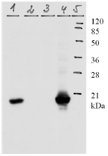
Western blot analysis of HPV-11 E7 protein. WB was carried out with 8G6 Mab using the lysates of U20S and Hela cells. Lines 1 and 4 are according cells transfected with plasmid expressing HPV-11 E7 protein, Lines 2 and 3 non-transfected U2OS and Hela cells. Line 5. Protein size marker.
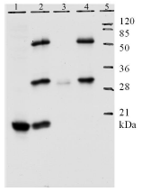
IP analysis of HPV-11 E7 protein. Line 1. shows detection of E7 protein by mAB 8G6 in lysate of U20S cells. Line 2. the lower band is precipitated E7, upper bands are light and heavy chain of Mab. Line 3. precipitation without mAB 8G6 Line 4. Nontransfected U2OS cells. Line 5. Protein size marker.
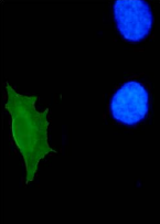
IF analysis of HPV-11 E7 protein in U2OS cells. IF was carried out with U2OS cells transfected with plasmid expressing HPV-11 E7 protein. All the nuclei were stained by DAPI. Green color represents nucleuslocalized HPV-11 E7 detected by Mab 9H5. IF analysis of E7 protein show similar result also in HeLa cells.
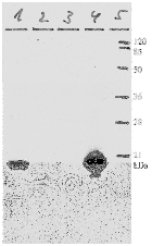
Western blot analysis of HPV-11 E7 protein. WB was carried out with 9H5 Mab using the lysates of U20S and Hela cells. Lines 1 and 4 are according cells transfected with plasmid expressing HPV-11 E7 protein, lines 2 and 3 non-transfected U2OS and Hela cells. Line 5. Protein size marker.
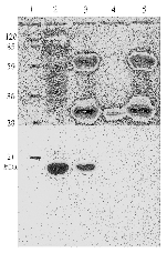
IP analysis of HPV-11 E7 protein. Line 2 detection of E7 protein by Mab 9H5 in lysate of U20S cells. Line 3 the lower band is precipitated E7, upper bands are light and heavy chain of Mab. Line 4 is precipitation without mab 8G6 and Line 5. lane shows IP experiment with plain U2OS cells. Line 1. Protein size marker.
Primary Antibodies
| Catalog No. | Host | Iso. | Clone | Pres. | React. | Applications | |
|---|---|---|---|---|---|---|---|
| 3HP11-711-13 | HPV11 E7 Protein antibody |
||||||
 |
Mouse | IgG1 | 711-13 | Purified | E, E(detection), WB |
1 mg /
€370.00
|
|
| HyTest Ltd. | |||||||

