KIAA1609 antibody
Principal name
KIAA1609 antibody
Alternative names for KIAA1609 antibody
TLD domain-containing protein KIAA1609
Ncbi ID
Available reactivities
Available hosts
Available applications
Western blot / Immunoblot (WB), Paraffin Sections (P), Immunocytochemistry/Immunofluorescence (ICC/IF), Flow Cytometry (F)
Background of KIAA1609 antibody
- Kit Component:
- - KN317608G1, 4632415K11Rik gRNA vector 1 in pCas-Guide vector
- KN317608G2, 4632415K11Rik gRNA vector 2 in pCas-Guide vector
- KN317608D, donor vector containing left and right homologous arms and Luciferase-Puro functional cassette
- GE100003, scramble sequence in pCas-Guide vector - CRISPR Manual:
- CRISPR/CAS9 Genome Editing Manual
General readings
Rothenberg, M.E., et al. Nat. Genet. 42(4):289-291(2010)
Venkatesan, K., et al. Nat. Methods 6(1):83-90(2009)
Wang, A.G., et al. Biochem. Biophys. Res. Commun. 345(3):1022-1032(2006)
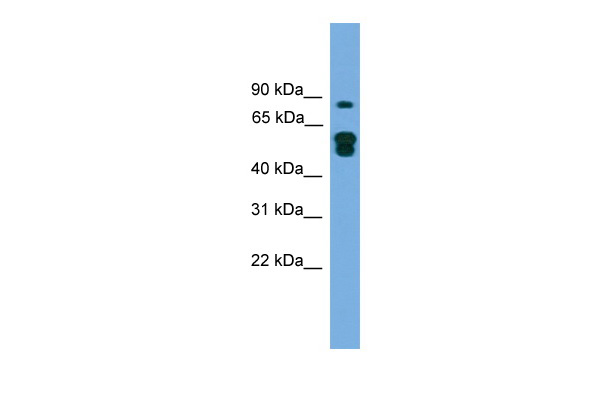
Transfected 293T; WB Suggested Anti-KIAA1609 Antibody Titration: 0.2-1 ug/ml. ELISA Titer: 1:1562500. Positive Control: Transfected 293T; KIAA1609 antibody - N-terminal region (ARP57503_P050) in Transfected 293T cells using Western Blot
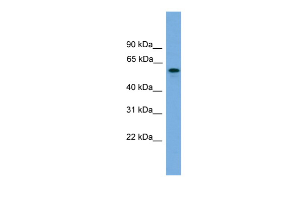
Transfected 293T; WB Suggested Anti-KIAA1609 Antibody Titration: 0.2-1 ug/ml. ELISA Titer: 1:1562500. Positive Control: Transfected 293T; KIAA1609 antibody - middle region (ARP57504_P050) in Transfected 293T cells using Western Blot

Hum. Fetal Brain; Host:Rabbit. Target Name:KIAA1609. Sample Tissue:Human Fetal Brain. Antibody Dilution: 1.0ug/ml.; KIAA1609 antibody - middle region (ARP57504_P050) in Hum. Fetal Brain cells using Western Blot
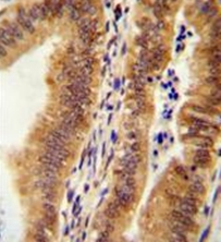
Immunohistochemistry analysis in formalin fixed and paraffin embedded human uterus tissue reacted with KIAA1609 Antibody (C-term) Cat.-No AP52284PU-N, which was peroxidase conjugated to the secondary antibody and followed by AB staining.
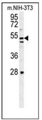
Western blot analysis of KIAA1609 Antibody (C-term) Cat.-No AP52284PU-N in mouse NIH-3T3 cell line lysates (35ug/lane).
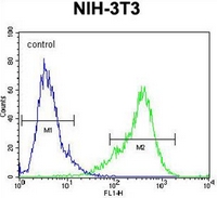
Flow cytometric analysis of NIH-3T3 cells using KIAA1609 Antibody (C-term) Cat.-No AP52284PU-N (right histogram) compared to a negative control cell (left histogram). FITC-conjugated goat-anti-rabbit secondary antibodies were used for the analysis.
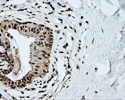
Immunohistochemical staining of paraffin-embedded breast tissue within the normal limits using anti-KIAA1609 mouse monoclonal antibody. (Heat-induced epitope retrieval by 10mM citric buffer, pH6.0, 100C for 10min, TA500962, Dilution 1:50)
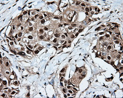
Immunohistochemical staining of paraffin-embedded Adenocarcinoma of breast tissue using anti-KIAA1609 mouse monoclonal antibody. (Heat-induced epitope retrieval by 10mM citric buffer, pH6.0, 100C for 10min, TA500962, Dilution 1:50)
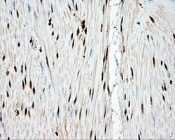
Immunohistochemical staining of paraffin-embedded colon tissue within the normal limits using anti-KIAA1609 mouse monoclonal antibody. (Heat-induced epitope retrieval by 10mM citric buffer, pH6.0, 100C for 10min, TA500962, Dilution 1:50)
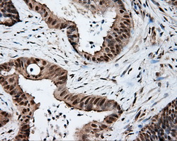
Immunohistochemical staining of paraffin-embedded Adenocarcinoma of colon tissue using anti-KIAA1609 mouse monoclonal antibody. (Heat-induced epitope retrieval by 10mM citric buffer, pH6.0, 100C for 10min, TA500962, Dilution 1:50)
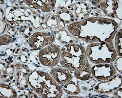
Immunohistochemical staining of paraffin-embedded Kidney tissue within the normal limits using anti-KIAA1609 mouse monoclonal antibody. (Heat-induced epitope retrieval by 10mM citric buffer, pH6.0, 100C for 10min, TA500962, Dilution 1:50)

Immunohistochemical staining of paraffin-embedded endometrium tissue within the normal limits using anti-KIAA1609 mouse monoclonal antibody. (Heat-induced epitope retrieval by 10mM citric buffer, pH6.0, 100C for 10min, TA500962, Dilution 1:50)

Immunohistochemical staining of paraffin-embedded liver tissue within the normal limits using anti-KIAA1609 mouse monoclonal antibody. (Heat-induced epitope retrieval by 10mM citric buffer, pH6.0, 100C for 10min, TA500962, Dilution 1:50)
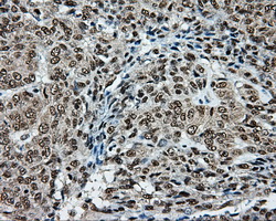
Immunohistochemical staining of paraffin-embedded Adenocarcinoma of endometrium tissue using anti-KIAA1609 mouse monoclonal antibody. (Heat-induced epitope retrieval by 10mM citric buffer, pH6.0, 100C for 10min, TA500962, Dilution 1:50)
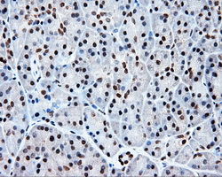
Immunohistochemical staining of paraffin-embedded pancreas tissue within the normal limits using anti-KIAA1609 mouse monoclonal antibody. (Heat-induced epitope retrieval by 10mM citric buffer, pH6.0, 100C for 10min, TA500962, Dilution 1:50)
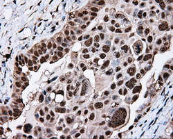
Immunohistochemical staining of paraffin-embedded Adenocarcinoma of ovary tissue using anti-KIAA1609 mouse monoclonal antibody. (Heat-induced epitope retrieval by 10mM citric buffer, pH6.0, 100C for 10min, TA500962, Dilution 1:50)
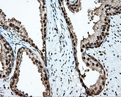
Immunohistochemical staining of paraffin-embedded prostate tissue within the normal limits using anti-KIAA1609 mouse monoclonal antibody. (Heat-induced epitope retrieval by 10mM citric buffer, pH6.0, 100C for 10min, TA500962, Dilution 1:50)

Immunohistochemical staining of paraffin-embedded Carcinoma of bladder tissue using anti-KIAA1609 mouse monoclonal antibody. (Heat-induced epitope retrieval by 10mM citric buffer, pH6.0, 100C for 10min, TA500962, Dilution 1:50)
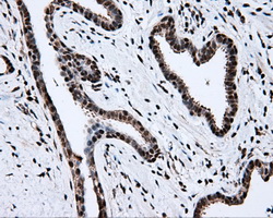
Immunohistochemical staining of paraffin-embedded Carcinoma of prostate tissue using anti-KIAA1609 mouse monoclonal antibody. (Heat-induced epitope retrieval by 10mM citric buffer, pH6.0, 100C for 10min, TA500962, Dilution 1:50)
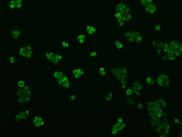
Immunofluorescent staining of HepG2 cells using anti-KIAA1609 mouse monoclonal antibody (TA500962).

HEK293T cells were transfected with the pCMV6-ENTRY control (Left lane) or pCMV6-ENTRY KIAA1609 (RC209287, Right lane) cDNA for 48 hrs and lysed. Equivalent amounts of cell lysates (5 ug per lane) were separated by SDS-PAGE and immunoblotted with anti-KIAA1609.
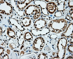
Immunohistochemical staining of paraffin-embedded Kidney tissue within the normal limits using anti-KIAA1609 mouse monoclonal antibody. (Heat-induced epitope retrieval by 10mM citric buffer, pH6.0, 100C for 10min, TA500963, Dilution 1:50)
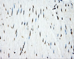
Immunohistochemical staining of paraffin-embedded colon tissue within the normal limits using anti-KIAA1609 mouse monoclonal antibody. (Heat-induced epitope retrieval by 10mM citric buffer, pH6.0, 100C for 10min, TA500963, Dilution 1:50)
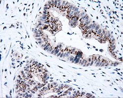
Immunohistochemical staining of paraffin-embedded Adenocarcinoma of colon tissue using anti-KIAA1609 mouse monoclonal antibody. (Heat-induced epitope retrieval by 10mM citric buffer, pH6.0, 100C for 10min, TA500963, Dilution 1:50)

Anti-KIAA1609 mouse monoclonal antibody (TA500963) immunofluorescent staining of COS7 cells transiently transfected by pCMV6-ENTRY KIAA1609(RC209287).
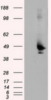
HEK293T cells were transfected with the pCMV6-ENTRY control (Left lane) or pCMV6-ENTRY KIAA1609 (RC209287, Right lane) cDNA for 48 hrs and lysed. Equivalent amounts of cell lysates (5 ug per lane) were separated by SDS-PAGE and immunoblotted with anti-KIAA1609.
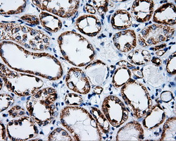
Immunohistochemical staining of paraffin-embedded Kidney tissue within the normal limits using anti-KIAA1609 mouse monoclonal antibody. (Heat-induced epitope retrieval by 10mM citric buffer, pH6.0, 100C for 10min, TA500980, Dilution 1:50)
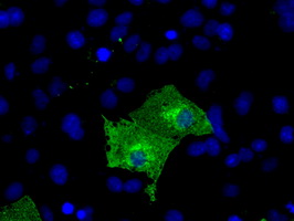
Anti-KIAA1609 mouse monoclonal antibody (TA500980) immunofluorescent staining of COS7 cells transiently transfected by pCMV6-ENTRY KIAA1609(RC209287).
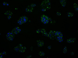
Immunofluorescent staining of HepG2 cells using anti-KIAA1609 mouse monoclonal antibody (TA500980).
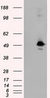
HEK293T cells were transfected with the pCMV6-ENTRY control (Left lane) or pCMV6-ENTRY KIAA1609 (RC209287, Right lane) cDNA for 48 hrs and lysed. Equivalent amounts of cell lysates (5 ug per lane) were separated by SDS-PAGE and immunoblotted with anti-KIAA1609.
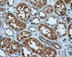
Immunohistochemical staining of paraffin-embedded Kidney tissue within the normal limits using anti-KIAA1609 mouse monoclonal antibody. (Heat-induced epitope retrieval by 10mM citric buffer, pH6.0, 100C for 10min, TA501008, Dilution 1:50)
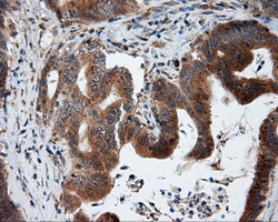
Immunohistochemical staining of paraffin-embedded Adenocarcinoma of colon tissue using anti-KIAA1609 mouse monoclonal antibody. (Heat-induced epitope retrieval by 10mM citric buffer, pH6.0, 100C for 10min, TA501008, Dilution 1:50)

Immunohistochemical staining of paraffin-embedded Carcinoma of liver tissue using anti-KIAA1609 mouse monoclonal antibody. (Heat-induced epitope retrieval by 10mM citric buffer, pH6.0, 100C for 10min, TA501008, Dilution 1:50)
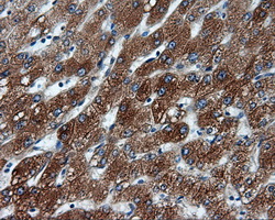
Immunohistochemical staining of paraffin-embedded liver tissue within the normal limits using anti-KIAA1609 mouse monoclonal antibody. (Heat-induced epitope retrieval by 10mM citric buffer, pH6.0, 100C for 10min, TA501008, Dilution 1:50)
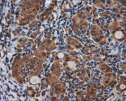
Immunohistochemical staining of paraffin-embedded Carcinoma of thyroid tissue using anti-KIAA1609 mouse monoclonal antibody. (Heat-induced epitope retrieval by 10mM citric buffer, pH6.0, 100C for 10min, TA501008, Dilution 1:50)
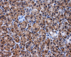
Immunohistochemical staining of paraffin-embedded pancreas tissue within the normal limits using anti-KIAA1609 mouse monoclonal antibody. (Heat-induced epitope retrieval by 10mM citric buffer, pH6.0, 100C for 10min, TA501008, Dilution 1:50)
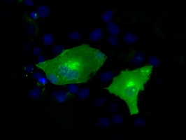
Anti-KIAA1609 mouse monoclonal antibody (TA501008) immunofluorescent staining of COS7 cells transiently transfected by pCMV6-ENTRY KIAA1609(RC209287).
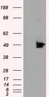
HEK293T cells were transfected with the pCMV6-ENTRY control (Left lane) or pCMV6-ENTRY KIAA1609 (RC209287, Right lane) cDNA for 48 hrs and lysed. Equivalent amounts of cell lysates (5 ug per lane) were separated by SDS-PAGE and immunoblotted with anti-KIAA1609.
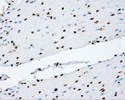
Immunohistochemical staining of paraffin-embedded colon tissue within the normal limits using anti-KIAA1609 mouse monoclonal antibody. (Heat-induced epitope retrieval by 10mM citric buffer, pH6.0, 100C for 10min, TA501037, Dilution 1:50)
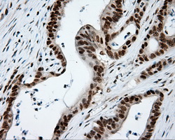
Immunohistochemical staining of paraffin-embedded Adenocarcinoma of colon tissue using anti-KIAA1609 mouse monoclonal antibody. (Heat-induced epitope retrieval by 10mM citric buffer, pH6.0, 100C for 10min, TA501037, Dilution 1:50)
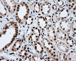
Immunohistochemical staining of paraffin-embedded Kidney tissue within the normal limits using anti-KIAA1609 mouse monoclonal antibody. (Heat-induced epitope retrieval by 10mM citric buffer, pH6.0, 100C for 10min, TA501037, Dilution 1:50)
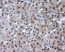
Immunohistochemical staining of paraffin-embedded liver tissue within the normal limits using anti-KIAA1609 mouse monoclonal antibody. (Heat-induced epitope retrieval by 10mM citric buffer, pH6.0, 100C for 10min, TA501037, Dilution 1:50)
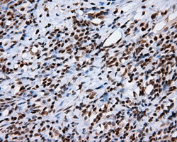
Immunohistochemical staining of paraffin-embedded Carcinoma of thyroid tissue using anti-KIAA1609 mouse monoclonal antibody. (Heat-induced epitope retrieval by 10mM citric buffer, pH6.0, 100C for 10min, TA501037, Dilution 1:50)
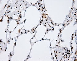
Immunohistochemical staining of paraffin-embedded lung tissue within the normal limits using anti-KIAA1609 mouse monoclonal antibody. (Heat-induced epitope retrieval by 10mM citric buffer, pH6.0, 100C for 10min, TA501037, Dilution 1:50)
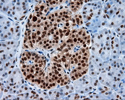
Immunohistochemical staining of paraffin-embedded pancreas tissue within the normal limits using anti-KIAA1609 mouse monoclonal antibody. (Heat-induced epitope retrieval by 10mM citric buffer, pH6.0, 100C for 10min, TA501037, Dilution 1:50)
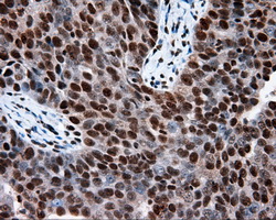
Immunohistochemical staining of paraffin-embedded Adenocarcinoma of ovary tissue using anti-KIAA1609 mouse monoclonal antibody. (Heat-induced epitope retrieval by 10mM citric buffer, pH6.0, 100C for 10min, TA501037, Dilution 1:50)

Immunohistochemical staining of paraffin-embedded endometrium tissue within the normal limits using anti-KIAA1609 mouse monoclonal antibody. (Heat-induced epitope retrieval by 10mM citric buffer, pH6.0, 100C for 10min, TA501037, Dilution 1:50)

Immunohistochemical staining of paraffin-embedded prostate tissue within the normal limits using anti-KIAA1609 mouse monoclonal antibody. (Heat-induced epitope retrieval by 10mM citric buffer, pH6.0, 100C for 10min, TA501037, Dilution 1:50)
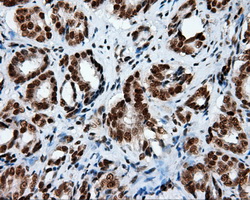
Immunohistochemical staining of paraffin-embedded Carcinoma of prostate tissue using anti-KIAA1609 mouse monoclonal antibody. (Heat-induced epitope retrieval by 10mM citric buffer, pH6.0, 100C for 10min, TA501037, Dilution 1:50)
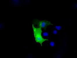
Anti-KIAA1609 mouse monoclonal antibody (TA501037) immunofluorescent staining of COS7 cells transiently transfected by pCMV6-ENTRY KIAA1609(RC209287).
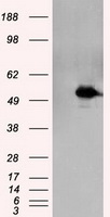
HEK293T cells were transfected with the pCMV6-ENTRY control (Left lane) or pCMV6-ENTRY KIAA1609 (RC209287, Right lane) cDNA for 48 hrs and lysed. Equivalent amounts of cell lysates (5 ug per lane) were separated by SDS-PAGE and immunoblotted with anti-KIAA1609.
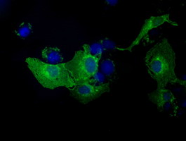
Anti-KIAA1609 mouse monoclonal antibody (TA501057) immunofluorescent staining of COS7 cells transiently transfected by pCMV6-ENTRY KIAA1609(RC209287).
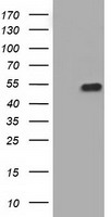
HEK293T cells were transfected with the pCMV6-ENTRY control (Left lane) or pCMV6-ENTRY KIAA1609 (RC209287, Right lane) cDNA for 48 hrs and lysed. Equivalent amounts of cell lysates (5 ug per lane) were separated by SDS-PAGE and immunoblotted with anti-KIAA1609.
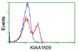
HEK293T cells transfected with either RC209287 overexpress plasmid(Red) or empty vector control plasmid(Blue) were immunostained by anti-KIAA1609 antibody(TA501057), and then analyzed by flow cytometry.
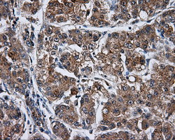
Immunohistochemical staining of paraffin-embedded Carcinoma of Human liver tissue using anti-KIAA1609 mouse monoclonal antibody. (Heat-induced epitope retrieval by 10mM citric buffer, pH6.0, 100C for 10min, TA501095)
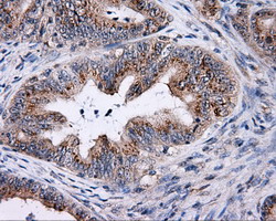
Immunohistochemical staining of paraffin-embedded Adenocarcinoma of Human colon tissue using anti-KIAA1609 mouse monoclonal antibody. (Heat-induced epitope retrieval by 10mM citric buffer, pH6.0, 100C for 10min, TA501095)
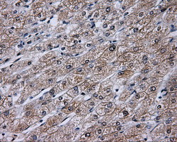
Immunohistochemical staining of paraffin-embedded Human liver tissue within the normal limits using anti-KIAA1609 mouse monoclonal antibody. (Heat-induced epitope retrieval by 10mM citric buffer, pH6.0, 100C for 10min, TA501095)
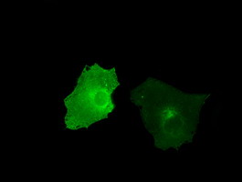
Anti-KIAA1609 mouse monoclonal antibody (TA501095) immunofluorescent staining of COS7 cells transiently transfected by pCMV6-ENTRY KIAA1609(RC209287).
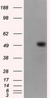
HEK293T cells were transfected with the pCMV6-ENTRY control (Left lane) or pCMV6-ENTRY KIAA1609 (RC209287, Right lane) cDNA for 48 hrs and lysed. Equivalent amounts of cell lysates (5 ug per lane) were separated by SDS-PAGE and immunoblotted with anti-KIAA1609.
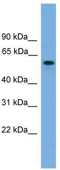
WB Suggested Anti-KIAA1609 Antibody Titration: 0.2-1 ug/ml; ELISA Titer: 1:1562500; Positive Control: Transfected 293T

Host:Rabbit; Target Name:KIAA1609; Sample Tissue:Human Fetal Brain; Antibody Dilution: 1.0 ug/ml
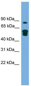
WB Suggested Anti-KIAA1609 Antibody Titration: 0.2-1 ug/ml; ELISA Titer: 1:1562500; Positive Control: Transfected 293T
Primary Antibodies
| Catalog No. | Host | Iso. | Clone | Pres. | React. | Applications | |
|---|---|---|---|---|---|---|---|
| TA501057 | KIAA1609 antibody |
||||||
| anti-KIAA1609 mouse monoclonal antibody, clone OTI3B1 (formerly 3B1) | |||||||
 |
Mouse | IgG2a | OTI3B1 | Purified | Hu | F, ICC/IF, WB |
0.1 ml /
€360.00
|
| OriGene Technologies, Inc. | |||||||
 +2 additional images +2 additional images |
|
||||||