MAFK antibody
Principal name
MAFK antibody
Alternative names for MAFK antibody
Transcription factor MafK, v-maf musculoaponeurotic fibrosarcoma oncogene homolog K
Ncbi ID
Available reactivities
Available hosts
Available applications
Enzyme Immunoassay (E), Western blot / Immunoblot (WB)
Background of MAFK antibody
The developmentally regulated expression of the globin genes depends on upstream regulatory elements termed locus control regions (LCRs). LCRs are associated with powerful enhancer activity that is mediated by the transcription factor NFE2 (nuclear factor erythroid-2). NFE2 recognition sites are also present in the gene promoters of 2 heme biosynthetic enzymes, porphobilinogen deamise (PBGD; MIM 609806) and ferrochelatase (FECH; MIM 612386). NFE2 D-binding activity consists of a heterodimer containing an 18-kD Maf protein (MafF, MafG (MIM 602020), or MafK) and p45 (MIM 601490). Both subunits are members of the activator protein-1 superfamily of basic leucine zipper (bZIP) proteins (see MIM 165160). Maf homodimers suppress transcription at NFE2 sites.[supplied by OMIM, Nov 2008]. Publication Note: This RefSeq record includes a subset of the publications that are available for this gene. Please see the Gene record to access additiol publications. ##Evidence-Data-START## Transcript exon combition :: AF059194.1, CT002504.1 [ECO:0000332] Rseq introns :: single sample supports all introns ERS025081, ERS025082 [ECO:0000348] ##Evidence-Data-END## COMPLETENESS: complete on the 3' end.
General readings
Iwata T, et al. (1998) Cytogenet Cell Genet 82 (1–2): 88–90.Du MJ, et al. (2008) Int J Biochem Cell Biol. 40(8):1481-93.

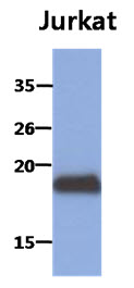
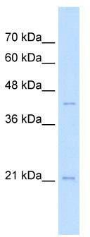
Primary Antibodies
| Catalog No. | Host | Iso. | Clone | Pres. | React. | Applications | |
|---|---|---|---|---|---|---|---|
| AM50032PU-N | MAFK antibody |
||||||
 |
Mouse | IgG2a | AT2F7 | Purified | Hu | E, WB |
0.1 ml /
€330.00
|
| OriGene Technologies GmbH | |||||||
| AM50032PU-S | MAFK antibody |
||||||
|
Mouse | IgG2a | AT2F7 | Purified | Hu | E, WB |
50 µl /
€220.00
|
| OriGene Technologies GmbH | |||||||

