MAGE-4 antibody
Principal name
MAGE-4 antibody
Alternative names for MAGE-4 antibody
MAGEA4, MAGE4, MAGE 4, Melanoma-associated antigen 4, Cancer/testis antigen 1.4
SwissProt ID
P43358 (Human)
Gene ID
4103 (MAGEA4)
Ncbi ID
NP_001011548.1, NP_001011548, 9606, AAH17723, NP_001011549, NP_001011550, NM_001011549, NM_001011548, NM_002362, NM_001011550
Available reactivities
Available hosts
Available applications
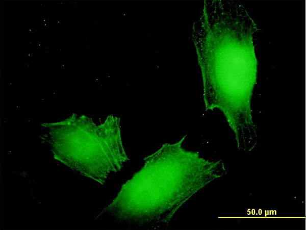
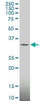
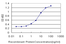
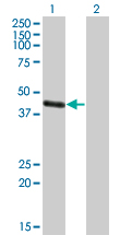
Enzyme Immunoassay (E), Immunocytochemistry/Immunofluorescence (ICC/IF), Western blot / Immunoblot (WB), ELISA (detection) (E(detection)), Paraffin Sections (P), Immunoprecipitation (IP)
Background of MAGE-4 antibody
MAGEA4 is a member of the MAGEA family. The members of this family are proteins with 50 to 80% sequence identity to each other. The promoters and first exons of the MAGEA genes show considerable variability, suggesting that the existence of this gene family enables the same function to be expressed under different transcriptional controls. The MAGEA genes are clustered at chromosomal location Xq28. They have been implicated in some hereditary disorders, such as dyskeratosis congenita.This gene is a member of the MAGEA gene family. The members of this family encode proteins with 50 to 80% sequence identity to each other. The promoters and first exons of the MAGEA genes show considerable variability, suggesting that the existence of this gene family enables the same function to be expressed under different transcriptional controls. The MAGEA genes are clustered at chromosomal location Xq28. They have been implicated in some hereditary disorders, such as dyskeratosis congenita. At least four variants encoding the same protein have been found for this gene.
General readings
Fradet Y., et al. (2009) Int J Cancer. 125(6):1365-71.
Nishimura T., et al. (2009) Br J Cancer. 100(7):1135-43.
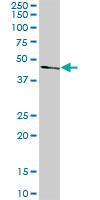
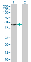
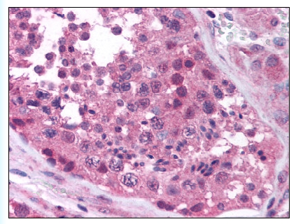
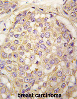

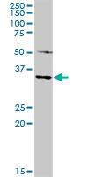
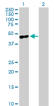
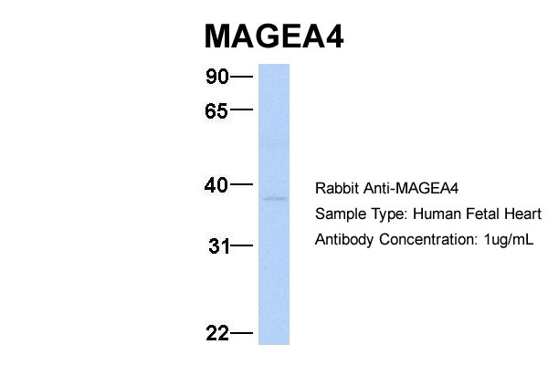
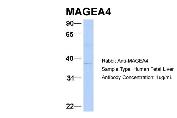

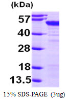
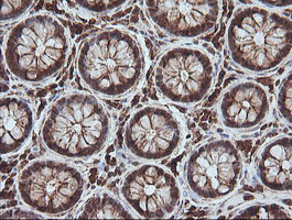
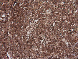
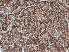

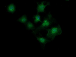

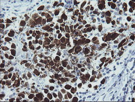
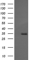

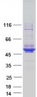

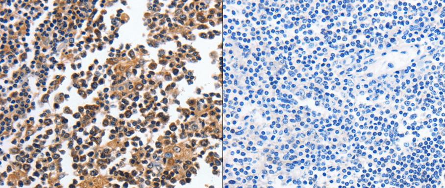
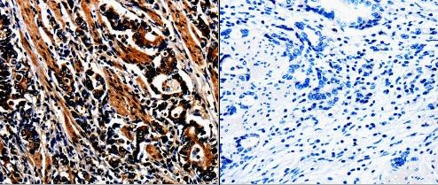
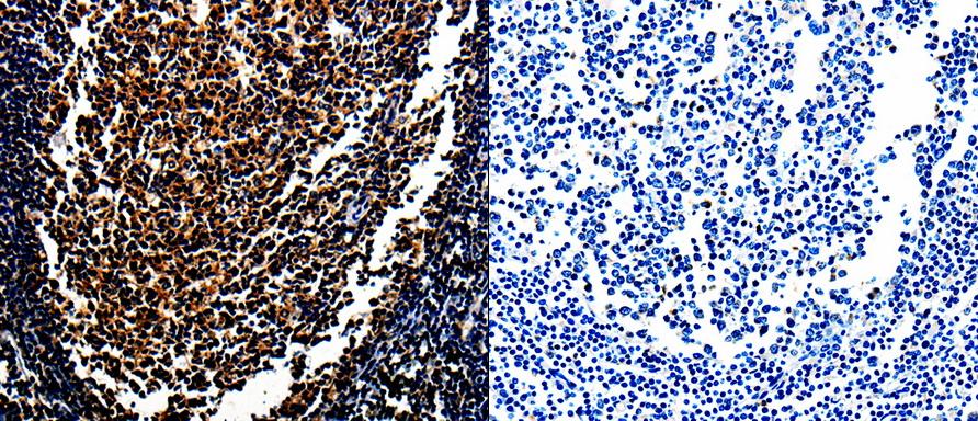
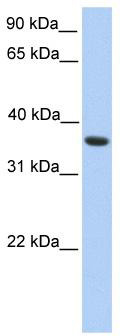
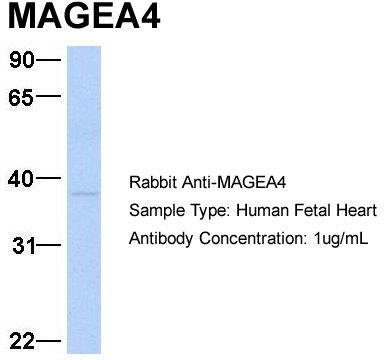

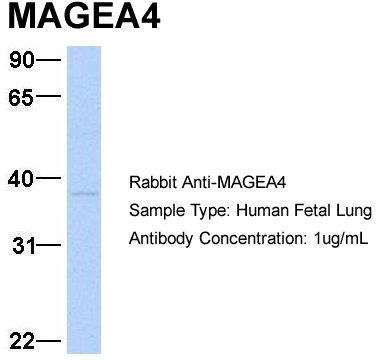
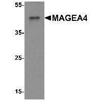
Primary Antibodies
| Catalog No. | Host | Iso. | Clone | Pres. | React. | Applications | |
|---|---|---|---|---|---|---|---|
| TA323460 | MAGE-4 antibody |
||||||
| Anti-MAGEA4 Rabbit Polyclonal Antibody | |||||||
 |
Rabbit | IgG | Purified | Hu, Ms, Rt | E, P |
0.1 ml /
€335.00
|
|
| OriGene Technologies, Inc. | |||||||
 +1 additional image +1 additional image |
|||||||
| TA323461 | MAGE-4 antibody |
||||||
| Anti-MAGEA4 Rabbit Polyclonal Antibody | |||||||
 |
Rabbit | IgG | Purified | Hu, Ms, Rt | E, P |
0.1 ml /
€335.00
|
|
| OriGene Technologies, Inc. | |||||||
| TA349177 | MAGE-4 antibody |
||||||
| Rabbit Polyclonal Anti-MAGEA4 Antibody | |||||||
 |
Rabbit | IgG | Purified | Hu, Ms, Rt | WB |
0.1 mg /
€415.00
|
|
| OriGene Technologies, Inc. | |||||||
| AP13131PU-N | MAGE-4 (N-term) antibody |
||||||
 |
Rabbit | Ig | Purified | Hu | P, WB |
0.4 ml /
€415.00
|
|
| OriGene Technologies GmbH | |||||||
| +1 additional image |
|||||||