MAP-2 antibody
Principal name
MAP-2 antibody
Alternative names for MAP-2 antibody
Microtubule-associated protein 2, MAP2, Neuronal Marker
Gene ID
4133 (MAP2)
Available reactivities
Available hosts
Available applications
Frozen Sections (C), Paraffin Sections (P), Western blot / Immunoblot (WB), Immunocytochemistry/Immunofluorescence (ICC/IF), ELISA (detection) (E(detection)), Immunoprecipitation (IP), Enzyme Immunoassay (E)
Background of MAP-2 antibody
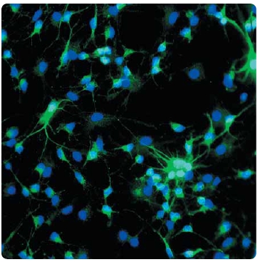
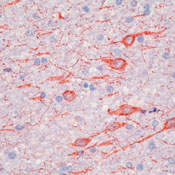
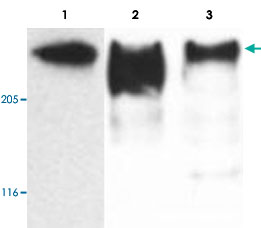
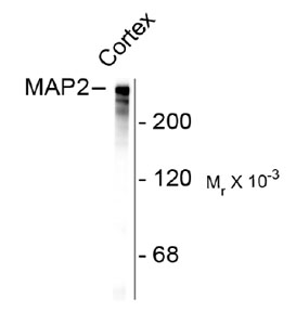
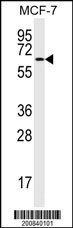
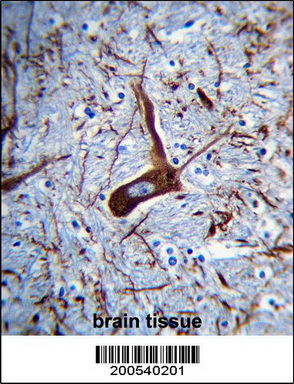
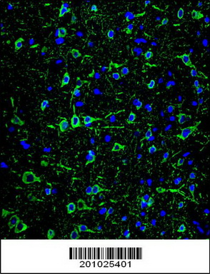
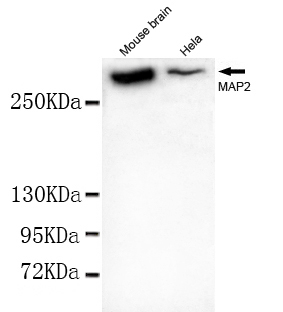
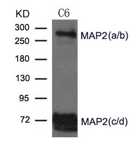
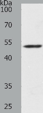
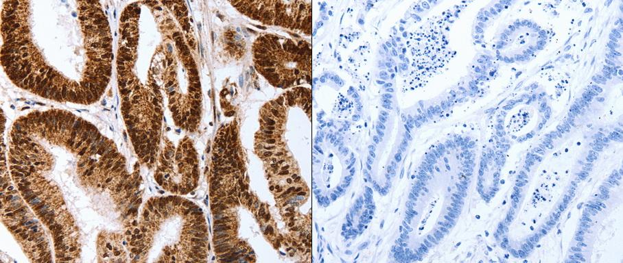
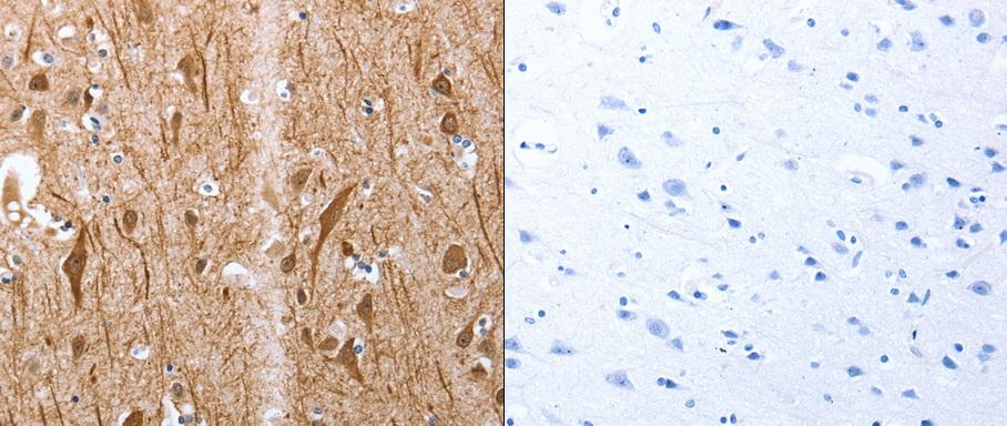
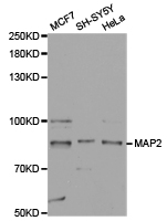
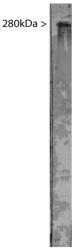
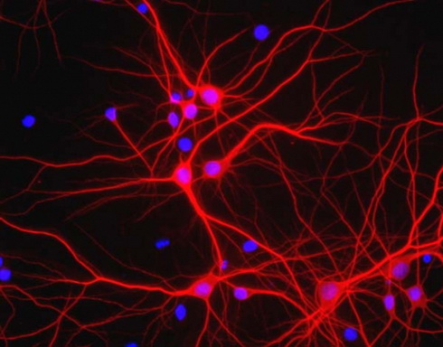
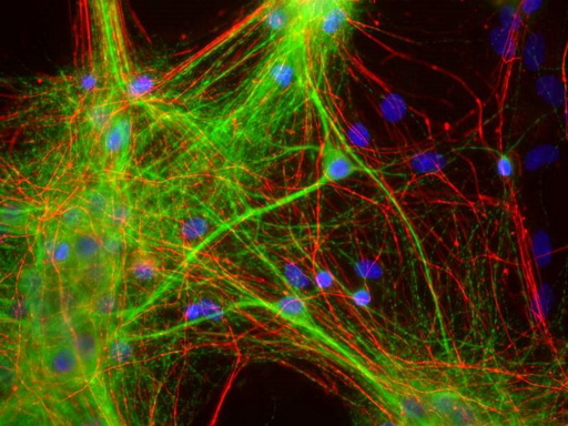
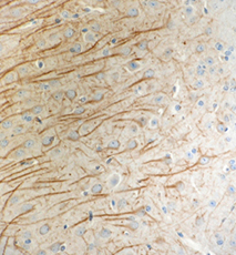
Microtubules are 25nm diameter protein rods found in most kinds of eukarytic cells. They are polymerized from a dimeric subunit made of one alpha subunit and one beta tubulin subunit. Microtubules are associated with a family of proteins called microtubule associated proteins (MAPs), which includes the protein tau and a group of proteins referred to as MAP1, MAP2, MAP3, MAP4 and MAP5. MAP2 is made up of two ~280kDa apparent molecular weight bands referred to as MAP2a and MAP2b. A third lower molecular weight form, usually called MAP2c, corresponds to a pair of protein bands running at ~70kDa on SDS-PAGE gels, and is expressed early in development. All these MAP2 forms are derived from a single gene by alterte transcription, and all share a C-termil sequence which includes either three or four microtubule binding peptide sequences, which are very similar to those found in the related microtubule binding protein tau. MAP2 isoforms are expressed only in neurol cells and specifically in the perikarya and dendrites of these cells. Antibodies to MAP2 are therefore excellent markers on neurol cells, their perikarya and neurol dendrites. In contrast tau is found predomintly in neurol axons.
General readings
1. Fontaine-Lenoir, V.; Chambraud, B.; Fellous, A.; David, S.; Duchossoy, Y.; Baulieu, E.-E.; Robel, P. :
Microtubule-associated protein 2 (MAP2) is a neurosteroid receptor. Proc. Nat. Acad. Sci. 103: 4711-4716, 2006.
2. Neve, R. L.; Harris, P.; Kosik, K. S.; Kurnit, D. M.; Donlon, T. A. : Identification of cDNA clones for the human microtubule-associated protein tau and chromosomal localization of the genes for tau and microtubule-associated protein 2. Molec. Brain Res. 1: 271-280, 1986.
3. Kalcheva, N.; Albala, J.; O'Guin, K.; Rubino, H.; Garner, C.; Shafit-Zagardo, B. : Genomic structure of human microtubule-associated protein 2 (MAP-2) and characterization of additional MAP-2 isoforms. Proc. Nat. Acad. Sci. 92: 10894-10898, 1995.








- Quick-Filters:
- Overnight Delivery (1)
Primary Antibodies
| Catalog No. | Host | Iso. | Clone | Pres. | React. | Applications | |
|---|---|---|---|---|---|---|---|
| CH22103-50 | MAP-2 antibody |
||||||
 |
Chicken | Serum | Hu, Ms, Prim, Rt | C, ICC/IF, WB |
50 µl /
€390.00
|
||
| Neuromics Antibodies | |||||||
 +1 additional image +1 additional image |
Top Selling product! |
||||||
| TA309162 | MAP-2 antibody |
||||||
| Chicken Anti-Microtubule Associated Protein 2 (MAP2) Antibody | |||||||
 |
Chicken | Ig | Ig Fraction | Bov, Hu, Ms, Rt | C, ICC/IF, WB |
0.1 ml /
€355.00
|
|
| OriGene Technologies, Inc. | |||||||
| TA323368 | MAP-2 antibody |
||||||
| Anti-Map2 Rabbit Polyclonal Antibody | |||||||
 |
Rabbit | IgG | Purified | Hu, Ms, Rt | E, P, WB |
0.1 ml /
€335.00
|
|
| OriGene Technologies, Inc. | |||||||
| +1 additional image |
|||||||
| TA323369 | MAP-2 antibody |
||||||
| Anti-Map2 Rabbit Polyclonal Antibody | |||||||
 |
Rabbit | IgG | Purified | Hu, Ms, Rt | E, P |
0.1 ml /
€335.00
|
|
| OriGene Technologies, Inc. | |||||||
| +1 additional image |
|||||||
| TA327383 | MAP-2 antibody |
||||||
| MAP2 Rabbit Polyclonal Antibody | |||||||
 |
Rabbit | IgG | Purified | Hu, Ms, Rt | WB |
0.1 mg /
€335.00
|
|
| OriGene Technologies, Inc. | |||||||
| TA336617 | MAP-2 antibody |
||||||
| Chicken Polyclonal MAP2 Antibody | |||||||
![Western Blot: MAP2 Antibody [TA336617] - Western blot analysis of MAP2 expression in bovine brain extract using TA336617.](https://m1.acris-antibodies.com/catalog/product/cache/1/small_image/50x50/9df78eab33525d08d6e5fb8d27136e95/OriGene/NB300-213-1-W.jpg) |
Chicken | IgY | Purified | Bov, Hu, Ms, Rt | C, ICC/IF, WB |
50 µl /
€395.00
|
|
| OriGene Technologies, Inc. | |||||||
| +1 additional image |
|||||||
| TA347836 | MAP-2 antibody |
||||||
| Rabbit Polyclonal Anti-MAP2 Antibody | |||||||
 |
Rabbit | IgG | Purified | Hu, Ms, Rt | WB |
0.1 mg /
€335.00
|
|
| OriGene Technologies, Inc. | |||||||
| TA347897 | MAP-2 antibody |
||||||
| Rabbit Polyclonal Anti-MAP 2 Antibody | |||||||
 |
Rabbit | IgG | Purified | Hu, Ms, Rt | WB |
0.1 mg /
€335.00
|
|
| OriGene Technologies, Inc. | |||||||
| AP11882PU-N | MAP-2 (C-term) antibody |
||||||
 |
Rabbit | Ig | Purified | Hu | P, WB |
0.4 ml /
€415.00
|
|
| OriGene Technologies GmbH | |||||||
| +2 additional images |
|||||||
| AP20610PU-N | MAP-2 antibody |
||||||
 |
Rabbit | Aff - Purified | Hu, Ms, Rt | P, WB |
0.1 mg /
€370.00
|
||
| OriGene Technologies GmbH | |||||||
| +1 additional image |
|||||||
| AP31808PU-N | MAP-2 antibody |
||||||
 |
Chicken | Ig | Aff - Purified | Hu, Ms | ICC/IF, P, WB |
0.2 mg /
€550.00
|
|
| OriGene Technologies GmbH | |||||||
| CH23032-100 | MAP-2 antibody |
||||||
 |
Chicken | Aff - Purified | Hu, Ms | C, ICC/IF |
0.1 ml /
€180.00
|
||
| Neuromics Antibodies | |||||||