MARK1 antibody
Principal name
MARK1 antibody
Alternative names for MARK1 antibody
Serine/threonine-protein kinase MARK1, MARK, MAP/Microtubule Affinity-Regulating Kinase 1
Ncbi ID
9606, 10090, NP_061120.3, NM_018650, AAF72103.1, NP_663490.2, NM_145515, NM_053947
Available reactivities
Available hosts
Available applications
Enzyme Immunoassay (E), Western blot / Immunoblot (WB), Paraffin Sections (P), Immunocytochemistry/Immunofluorescence (ICC/IF)
Background of MARK1 antibody
- Kit Component:
- - KN309797G1, Mark1 gRNA vector 1 in pCas-Guide vector
- KN309797G2, Mark1 gRNA vector 2 in pCas-Guide vector
- KN309797D, donor vector containing left and right homologous arms and Luciferase-Puro functional cassette
- GE100003, scramble sequence in pCas-Guide vector - CRISPR Manual:
- CRISPR/CAS9 Genome Editing Manual
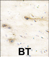
Formalin-fixed and paraffin-embedded human brain tissue reacted with MARK1 polyclonal antibody ( Cat # PAB2698 ) , which was peroxidase-conjugated to the secondary antibody, followed by DAB staining. This data demonstrates the use of this antibody for immunohistochemistry ; clinical relevance has not been evaluated.
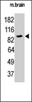
The MARK1 polyclonal antibody ( Cat # PAB2698 ) is used in Western blot to detect MARK1 in P7 mouse whole brain lysate ( 60 µg ) . 1 : 250 dilution of anti-MARK1 polyclonal antibody was used. Data and protocol kindly provided by DR. Shengli Zhao, M.D., Ph.D. of Duke University Medical Center.
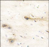
Formalin-fixed and paraffin-embedded human brain tissue reacted with MARK1 Antibody (N-term), which was peroxidase-conjugated to the secondary antibody, followed by DAB staining.
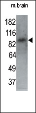
The anti-MARK1 N-term Pab is used in Western blot to detect MARK1 in P7 mouse whole brain lysate (60 ug). 1:250 dilution of anti-MARK1 pab was used.
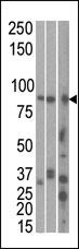
The anti-MARK1 C-term Pab is used in Western blot to detect MARK1 in, from left to right, Hela, T47D, and mouse brain cell line/ tissue lysate.
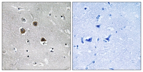

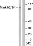
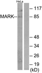

Immunohistochemical analysis of MARK staining in human lung cancer formalin fixed paraffin embedded tissue section. The section was pre-treated using heat mediated antigen retrieval with sodium citrate buffer (pH 6.0). The section was then incubated with the antibody at room temperature and detected using an HRP conjugated compact polymer system. DAB was used as the chromogen. The section was then counterstained with haematoxylin and mounted with DPX.
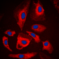
Immunofluorescent analysis of MARK staining in A549 cells. Formalin-fixed cells were permeabilized with 0.1% Triton X-100 in TBS for 5-10 minutes and blocked with 3% BSA-PBS for 30 minutes at room temperature. Cells were probed with the primary antibody in 3% BSA-PBS and incubated overnight at 4 C in a humidified chamber. Cells were washed with PBST and incubated with a DyLight 594-conjugated secondary antibody (red) in PBS at room temperature in the dark. DAPI was used to stain the cell nuclei (blue).

Western blot analysis of MARK expression in A549 (A); HeLa (B) whole cell lysates.
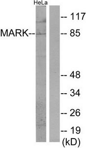
Western blot analysis of extracts from HeLa cells, using MARK antibody.The lane on the right is treated with the synthesized peptide.

Anti-MARK1 antibody IHC of human brain, cerebellum. Immunohistochemistry of formalin-fixed, paraffin-embedded tissue after heat-induced antigen retrieval.
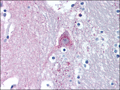
Brain, caduate: Formalin-Fixed Paraffin-Embedded (FFPE)
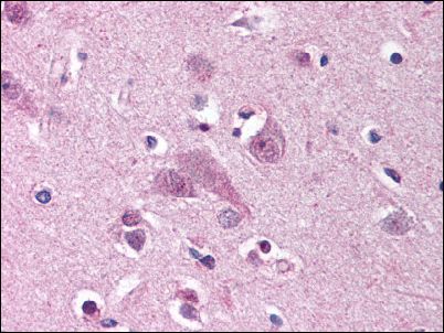
Brain, cortex: Formalin-Fixed Paraffin-Embedded (FFPE)

Gel: 6%SDS-PAGE, Lysate: 40 ug, Lane: Mouse brain tissue, Primary antibody: (MARK1 Antibody) at dilution 1/300, Secondary antibody: Goat anti rabbit IgG at 1/8000 dilution, Exposure time: 30 seconds
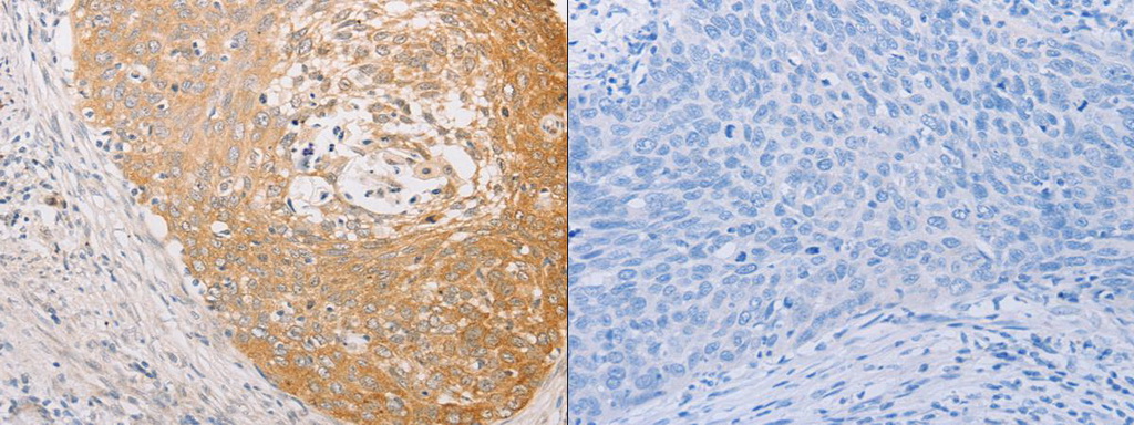
The image on the left is immunohistochemistry of paraffin-embedded Human cervical cancer tissue using (MARK1 Antibody) at dilution 1/30, on the right is treated with fusion protein. (Original magnification: ×200)
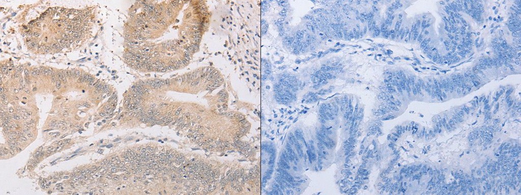
The image on the left is immunohistochemistry of paraffin-embedded Human colon cancer tissue using (MARK1 Antibody) at dilution 1/30, on the right is treated with fusion protein. (Original magnification: ×200)
Primary Antibodies
| Catalog No. | Host | Iso. | Clone | Pres. | React. | Applications | |
|---|---|---|---|---|---|---|---|
| TA314175 | MARK1 antibody |
||||||
| Rabbit polyclonal anti-MARK antibody | |||||||
 |
Rabbit | IgG | Purified | Hu, Ms, Rt | E, WB |
0.1 ml /
€335.00
|
|
| OriGene Technologies, Inc. | |||||||
| TA341169 | MARK1 antibody |
||||||
| Rabbit Polyclonal Anti-MARK1 Antibody (Internal) | |||||||
 |
Rabbit | Purified | Bov, Can, Eq, Hu |
50 µg /
€425.00
|
|||
| OriGene Technologies, Inc. | |||||||
| TA341195 | MARK1 (N-term) antibody |
||||||
| Rabbit Polyclonal Anti-MARK1 Antibody (N-Terminus) | |||||||
 |
Rabbit | Aff - Purified | Hu | P |
50 µg /
€425.00
|
||
| OriGene Technologies, Inc. | |||||||
| TA341196 | MARK1 (N-term) antibody |
||||||
| Rabbit Polyclonal Anti-MARK1 Antibody (N-Terminus) | |||||||
|
Rabbit | Aff - Purified | Hu | P |
50 µg /
€425.00
|
||
| OriGene Technologies, Inc. | |||||||
| TA349587 | MARK1 antibody |
||||||
| Rabbit Polyclonal Anti-MARK1 Antibody | |||||||
 |
Rabbit | IgG | Purified | Hu, Ms, Rt | E, P, WB |
0.1 ml /
€335.00
|
|
| OriGene Technologies, Inc. | |||||||
 +2 additional images +2 additional images |
|||||||
| AP13782PU-N | MARK1 (N-term) antibody |
||||||
 |
Rabbit | Ig | Purified | Hu, Ms | P, WB |
0.4 ml /
€415.00
|
|
| OriGene Technologies GmbH | |||||||
| +1 additional image |
|||||||
| AP13783PU-N | MARK1 (C-term) antibody |
||||||
 |
Rabbit | Ig | Purified | Hu, Ms | WB |
0.4 ml /
€415.00
|
|
| OriGene Technologies GmbH | |||||||
Mouse Lenti ORF Particles
| Catalog No. | ||
|---|---|---|
| MR215783L2V | Lenti ORF particles, Mark1 (GFP-tagged) - Mouse MAP/microtubule affinity-regulating kinase 1 (Mark1), 200 uL, >10^7 TU/mL |
|
|
200 µl /
€1,600.00
|
|
| OriGene Technologies, Inc. | ||
| MR215783L1V | Lenti ORF particles, Mark1 (Myc-DDK-tagged) - Mouse MAP/microtubule affinity-regulating kinase 1 (Mark1), 200 uL, >10^7 TU/mL |
|
|
200 µl /
€1,440.00
|
|
| OriGene Technologies, Inc. | ||
Rat Lenti ORF Particles
| Catalog No. | ||
|---|---|---|
| RR206808L2V | Lenti ORF particles, Mark1 (GFP-tagged ORF) - Rat MAP/microtubule affinity-regulating kinase 1 (Mark1), 200 uL, >10^7 TU/mL |
|
|
200 µl /
€1,600.00
|
|
| OriGene Technologies, Inc. | ||
| RR206808L1V | Lenti ORF particles, Mark1 (Myc-DDK-tagged ORF) - Rat MAP/microtubule affinity-regulating kinase 1 (Mark1), 200 uL, >10^7 TU/mL |
|
|
200 µl /
€1,440.00
|
|
| OriGene Technologies, Inc. | ||