MARK3 antibody
Principal name
MARK3 antibody
Alternative names for MARK3 antibody
CTAK1, C-TAK1, MAP/microtubule affinity-regulating kinase 3, Serine/threonine-protein kinase p78, PAR-1, STK10, Cdc25C-associated protein kinase 1
Ncbi ID
NP_001122390, NP_001122390.1, NP_002367, 9606, NM_002376.4, NP_002367.4, NP_001122393, NM_021516, NM_001128921, NM_001128919, BC026445, NM_002376, NM_001128918, NM_130749
Available reactivities
Available hosts
Available applications
Immunoprecipitation (IP), Paraffin Sections (P), Western blot / Immunoblot (WB), Enzyme Immunoassay (E), ELISA (detection) (E(detection))
Background of MARK3 antibody
- Kit Component:
- - KN205758G1, MARK3 gRNA vector 1 in pCas-Guide vector
- KN205758G2, MARK3 gRNA vector 2 in pCas-Guide vector
- KN205758D, donor vector containing left and right homologous arms and Luciferase-Puro functional cassette
- GE100003, scramble sequence in pCas-Guide vector - CRISPR Manual:
- CRISPR/CAS9 Genome Editing Manual
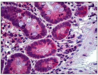
Human Small Intestine: Formalin-Fixed, Paraffin-Embedded (FFPE)
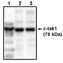
Western blot analysis using c-tak1 antibody on His-tagged c-tak1 (1), RKO cell lysate (2) and HCT116 cell lysate (3).

Mouse Brain; . Sample Type: Lane1 = 10ug mouse brain extract; Lane 2 = 50ug mouse brain extract. Primary Antibody Dilution: Anti-MARK3 1:2500. Submitted By: Dr. Veronique Dorval, Laval University.; MARK3 antibody - middle region (ARP56343_P050) in Mouse Brain cells using Western Blot
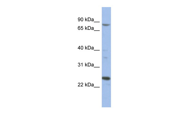
Human 293T; WB Suggested Anti-MARK3 Antibody Titration: 0.2-1 ug/ml. ELISA Titer: 1:312500. Positive Control: 293T cell lysate. MARK3 is supported by BioGPS gene expression data to be expressed in HEK293T.; MARK3 antibody - middle region (ARP56343_P050) in Human 293T cells using Western Blot

Western Blot analysis of MARK3 expression in transfected 293T cell line (H00004140-T01) by MARK3 MaxPab polyclonal antibody.Lane 1: MARK3 transfected lysate(80.19 KDa).Lane 2: Non-transfected lysate.
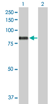
Western Blot analysis of MARK3 expression in transfected 293T cell line (H00004140-T01) by MARK3 MaxPab polyclonal antibody.Lane 1: MARK3 transfected lysate(80.19 KDa).Lane 2: Non-transfected lysate.
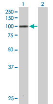
Western Blot analysis of MARK3 expression in transfected 293T cell line (H00004140-T02) by MARK3 MaxPab polyclonal antibody.Lane 1: MARK3 transfected lysate(81.40 KDa).Lane 2: Non-transfected lysate.
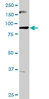
MARK3 MaxPab rabbit polyclonal antibody. Western Blot analysis of MARK3 expression in HeLa.
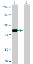
Western Blot analysis of MARK3 expression in transfected 293T cell line by MARK3 monoclonal antibody (M01), clone 1A10.Lane 1: MARK3 transfected lysate(81.4 KDa).Lane 2: Non-transfected lysate.
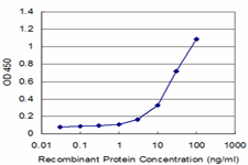
Detection limit for recombinant GST tagged MARK3 is approximately 3ng/ml as a capture antibody.
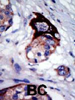
Formalin-fixed and paraffin-embedded human cancer tissue reacted with MARK3 polyclonal antibody ( Cat # PAB2052 ) , which was peroxidase-conjugated to the secondary antibody, followed by AEC staining.This data demonstrates the use of this antibody for immunohistochemistry ; clinical relevance has not been evaluated. BC = breast carcinoma.
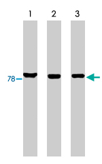
Western blot analysis using MARK3 polyclonal antibody on His-tagged c-tak1 (lane 1), RKO cell lysate (lane 2) and HCT-116 cell lysate (lane 3).
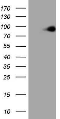
HEK293T cells were transfected with the pCMV6-ENTRY control (Left lane) or pCMV6-ENTRY MARK3 (RC205758, Right lane) cDNA for 48 hrs and lysed. Equivalent amounts of cell lysates (5 ug per lane) were separated by SDS-PAGE and immunoblotted with anti-MARK3.
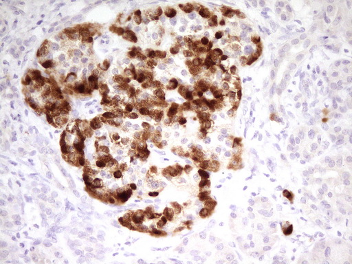
Immunohistochemical staining of paraffin-embedded Human pancreas tissue within the normal limits using anti-MARK3 mouse monoclonal antibody. (Heat-induced epitope retrieval by 1 mM EDTA in 10mM Tris, pH8.5, 120C for 3min, TA805764)

HEK293T cells were transfected with the pCMV6-ENTRY control (Left lane) or pCMV6-ENTRY MARK3 (RC205758, Right lane) cDNA for 48 hrs and lysed. Equivalent amounts of cell lysates (5 ug per lane) were separated by SDS-PAGE and immunoblotted with anti-MARK3.
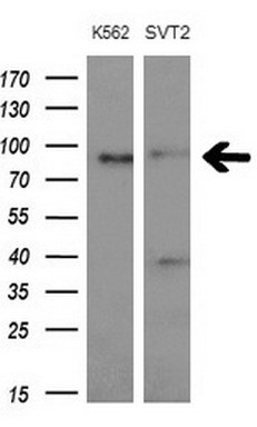
Western blot analysis of extracts (10ug) from 2 different cell lines by using anti- MARK3monoclonal antibody at 1:200 dilution.

Immunohistochemical staining of paraffin-embedded Carcinoma of Human liver tissue using anti-MARK3 mouse monoclonal antibody. (Heat-induced epitope retrieval by 1 mM EDTA in 10mM Tris, pH8.5, 120C for 3min, TA805790)

Immunohistochemical staining of paraffin-embedded Human liver tissue within the normal limits using anti-MARK3 mouse monoclonal antibody. (Heat-induced epitope retrieval by 1 mM EDTA in 10mM Tris, pH8.5, 120C for 3min, TA805790)

HEK293T cells were transfected with the pCMV6-ENTRY control (Left lane) or pCMV6-ENTRY MARK3 (RC205758, Right lane) cDNA for 48 hrs and lysed. Equivalent amounts of cell lysates (5 ug per lane) were separated by SDS-PAGE and immunoblotted with anti-MARK3.
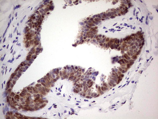
Immunohistochemical staining of paraffin-embedded Human breast tissue within the normal limits using anti-MARK3 mouse monoclonal antibody. (Heat-induced epitope retrieval by 1 mM EDTA in 10mM Tris, pH8.5, 120C for 3min, TA805880)

HEK293T cells were transfected with the pCMV6-ENTRY control (Left lane) or pCMV6-ENTRY MARK3 (RC205758, Right lane) cDNA for 48 hrs and lysed. Equivalent amounts of cell lysates (5 ug per lane) were separated by SDS-PAGE and immunoblotted with anti-MARK3.
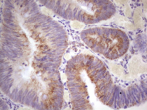
Immunohistochemical staining of paraffin-embedded Adenocarcinoma of Human colon tissue using anti-MARK3 mouse monoclonal antibody. (TA805881; heat-induced epitope retrieval by 1 mM EDTA in 10mM Tris, pH8.5, 120C for 3min)
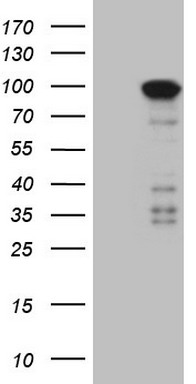
HEK293T cells were transfected with the pCMV6-ENTRY control (Left lane) or pCMV6-ENTRY MARK3 (RC205758, Right lane) cDNA for 48 hrs and lysed. Equivalent amounts of cell lysates (5 ug per lane) were separated by SDS-PAGE and immunoblotted with anti-MARK3.
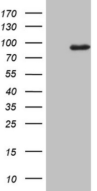
HEK293T cells were transfected with the pCMV6-ENTRY control (Left lane) or pCMV6-ENTRY MARK3 (RC205758, Right lane) cDNA for 48 hrs and lysed. Equivalent amounts of cell lysates (5 ug per lane) were separated by SDS-PAGE and immunoblotted with anti-MARK3.
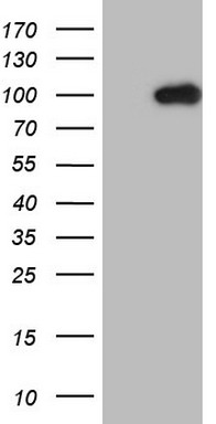
HEK293T cells were transfected with the pCMV6-ENTRY control (Left lane) or pCMV6-ENTRY MARK3 (RC205758, Right lane) cDNA for 48 hrs and lysed. Equivalent amounts of cell lysates (5 ug per lane) were separated by SDS-PAGE and immunoblotted with anti-MARK3.
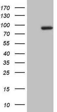
HEK293T cells were transfected with the pCMV6-ENTRY control (Left lane) or pCMV6-ENTRY MARK3 (RC205758, Right lane) cDNA for 48 hrs and lysed. Equivalent amounts of cell lysates (5 ug per lane) were separated by SDS-PAGE and immunoblotted with anti-MARK3.
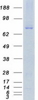

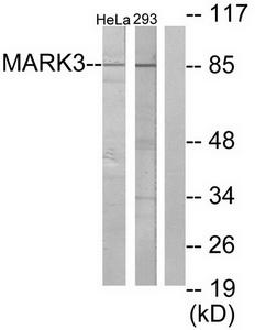
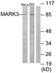
Western blot analysis of extracts from HeLa cells and 293 cells, using MARK3 antibody.The lane on the right is treated with the synthesized peptide.
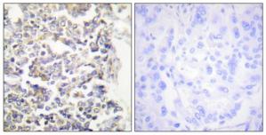
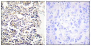
Immunohistochemistry analysis of paraffin-embedded human lung carcinoma tissue using MARK3 antibody.The picture on the right is treated with the synthesized peptide.
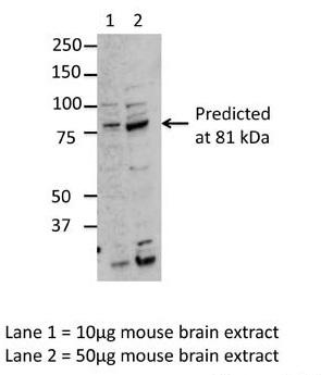
Sample Type: Lane1 = 10ug mouse brain extract; Lane 2 = 50ug mouse brain extract; Primary Antibody Dilution: Anti-MARK3 1:2500; Submitted By: Dr. Veronique Dorval, Laval University
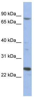
WB Suggested Anti-MARK3 Antibody Titration: 0.2-1 ug/ml; ELISA Titer: 1:312500; Positive Control: 293T cell lysateMARK3 is supported by BioGPS gene expression data to be expressed in HEK293T
Primary Antibodies
| Catalog No. | Host | Iso. | Clone | Pres. | React. | Applications | |
|---|---|---|---|---|---|---|---|
| TA311991 | MARK3 antibody |
||||||
| Rabbit polyclonal anti-MARK3 antibody | |||||||
 |
Rabbit | IgG | Purified | Hu, Ms, Rt | E, P, WB |
0.1 ml /
€335.00
|
|
| OriGene Technologies, Inc. | |||||||
 +1 additional image +1 additional image |
|||||||
| TA805763 | MARK3 antibody |
||||||
| MARK3 mouse monoclonal antibody, clone OTI2A4 (formerly 2A4) | |||||||
 |
Mouse | IgG1 | OTI2A4 | Purified | Hu | WB |
0.1 ml /
€360.00
|
| OriGene Technologies, Inc. | |||||||
|
|||||||
| TA805764 | MARK3 antibody |
||||||
| MARK3 mouse monoclonal antibody, clone OTI2G8 (formerly 2G8) | |||||||
 |
Mouse | IgG1 | OTI2G8 | Purified | Hu | P, WB |
0.1 ml /
€360.00
|
| OriGene Technologies, Inc. | |||||||
| +2 additional images |
|
||||||
| TA805765 | MARK3 antibody |
||||||
| MARK3 mouse monoclonal antibody, clone OTI2D3 (formerly 2D3) | |||||||
 |
Mouse | IgG1 | OTI2D3 | Purified | Hu | WB |
0.1 ml /
€360.00
|
| OriGene Technologies, Inc. | |||||||
|
|||||||
| TA805790 | MARK3 antibody |
||||||
| MARK3 mouse monoclonal antibody, clone OTI2D9 (formerly 2D9) | |||||||
 |
Mouse | IgG2a | OTI2D9 | Purified | Hu | P, WB |
0.1 ml /
€360.00
|
| OriGene Technologies, Inc. | |||||||
| +2 additional images |
|
||||||
| TA805880 | MARK3 antibody |
||||||
| MARK3 mouse monoclonal antibody, clone OTI3F6 (formerly 3F6) | |||||||
 |
Mouse | IgG1 | OTI3F6 | Purified | Hu | P, WB |
0.1 ml /
€360.00
|
| OriGene Technologies, Inc. | |||||||
| +1 additional image |
|
||||||
| TA805881 | MARK3 antibody |
||||||
| MARK3 mouse monoclonal antibody, clone OTI3E11 (formerly 3E11) | |||||||
 |
Mouse | IgG1 | OTI3E11 | Purified | Hu | P |
0.1 ml /
€360.00
|
| OriGene Technologies, Inc. | |||||||
|
|||||||
| TA805884 | MARK3 antibody |
||||||
| MARK3 mouse monoclonal antibody, clone OTI3D8 (formerly 3D8) | |||||||
 |
Mouse | IgG2b | OTI3D8 | Purified | Hu | WB |
0.1 ml /
€360.00
|
| OriGene Technologies, Inc. | |||||||
|
|||||||
| TA805954 | MARK3 antibody |
||||||
| MARK3 mouse monoclonal antibody, clone OTI4A3 (formerly 4A3) | |||||||
 |
Mouse | IgG1 | OTI4A3 | Purified | Hu | WB |
0.1 ml /
€360.00
|
| OriGene Technologies, Inc. | |||||||
|
|||||||
| TA805955 | MARK3 antibody |
||||||
| MARK3 mouse monoclonal antibody, clone OTI5E5 (formerly 5E5) | |||||||
 |
Mouse | IgG2b | OTI5E5 | Purified | Hu | WB |
0.1 ml /
€360.00
|
| OriGene Technologies, Inc. | |||||||
|
|||||||
| AP05080PU-N | MARK3 antibody |
||||||
 |
Sheep | IgG | Purified | Hu | IP, WB |
0.25 mg /
€400.00
|
|
| OriGene Technologies GmbH | |||||||

Proteins & Growth Factors
| Catalog No. | Species | Pres. | Purity | Source | |
|---|---|---|---|---|---|
| TP305758 | MARK3 (transcript variant 3) |
||||
| Recombinant protein of human MAP/microtubule affinity-regulating kinase 3 (MARK3), transcript variant 3 | |||||
 |
Human | > 80 % Preparation: Recombint protein was captured through anti-DDK affinity column followed by conventiol chromatography steps. Purity Detail: > 80% as determined by SDS-PAGE and Coomassie blue staining. |
HEK293 cells |
20 µg /
€748.00
|
|
| OriGene Technologies, Inc. | |||||
| TP760895 | MARK3 (full length, N-term HIS tag, transcript variant 5) |
||||
| Purified recombinant protein of Human MAP/microtubule affinity-regulating kinase 3 (MARK3), transcript variant 5, full length, with N-terminal HIS tag, expressed in E. coli, 50ug | |||||
 |
Human | > 80 % Preparation: . Purity Detail: > 80% as determined by SDS-PAGE and Coomassie blue staining. |
E. coli |
50 µg /
€205.00
|
|
| OriGene Technologies, Inc. | |||||
Mouse Lenti ORF Particles
| Catalog No. | ||
|---|---|---|
| MR206826L2V | Lenti ORF particles, Mark3 (GFP-tagged) - Mouse MAP/microtubule affinity-regulating kinase 3 (cDNA clone MGC:31426 IMAGE:4459439), 200 uL, >10^7 TU/mL |
|
 |
200 µl /
€1,240.00
|
|
| OriGene Technologies, Inc. | ||
| MR206826L1V | Lenti ORF particles, Mark3 (Myc-DDK-tagged) - Mouse MAP/microtubule affinity-regulating kinase 3 (cDNA clone MGC:31426 IMAGE:4459439), 200 uL, >10^7 TU/mL |
|
|
200 µl /
€1,110.00
|
|
| OriGene Technologies, Inc. | ||
Rat Lenti ORF Particles
| Catalog No. | ||
|---|---|---|
| RR213441L2V | Lenti ORF particles, Mark3 (GFP-tagged ORF) - Rat MAP/microtubule affinity-regulating kinase 3 (Mark3), 200 uL, >10^7 TU/mL |
|
|
200 µl /
€1,600.00
|
|
| OriGene Technologies, Inc. | ||
| RR213441L1V | Lenti ORF particles, Mark3 (Myc-DDK-tagged ORF) - Rat MAP/microtubule affinity-regulating kinase 3 (Mark3), 200 uL, >10^7 TU/mL |
|
|
200 µl /
€1,440.00
|
|
| OriGene Technologies, Inc. | ||