MAWBP antibody
Principal name
MAWBP antibody
Alternative names for MAWBP antibody
PBLD, Unknown protein 32 from 2D-page of liver tissue, Phenazine biosynthesis-like domain-containing protein
Ncbi ID
9606, NP_071412, NP_071412.2, NM_138530, NM_022129, NM_001033083
Available reactivities
Available hosts
Available applications
ELISA (detection) (E(detection)), Western blot / Immunoblot (WB), Flow Cytometry (F), Immunocytochemistry/Immunofluorescence (ICC/IF), Paraffin Sections (P)
Background of MAWBP antibody
- Kit Component:
- - KN202328G1, PBLD gRNA vector 1 in pCas-Guide vector
- KN202328G2, PBLD gRNA vector 2 in pCas-Guide vector
- KN202328D, donor vector containing left and right homologous arms and Luciferase-Puro functional cassette
- GE100003, scramble sequence in pCas-Guide vector - CRISPR Manual:
- CRISPR/CAS9 Genome Editing Manual
General readings
Mavrodi DV., et al. (2004) Acta Crystallogr D Biol Crystallogr. 60(1):184-6.
Parsons JF., et al. (2004) Biochemistry. 43(39):12427-35.
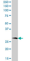
MAWBP MaxPab polyclonal antibody. Western Blot analysis of MAWBP expression in human liver.
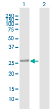
Western Blot analysis of PBLD expression in transfected 293T cell line (H00064081-T01) by PBLD MaxPab polyclonal antibody.Lane 1: MAWBP transfected lysate(31.68 KDa).Lane 2: Non-transfected lysate.
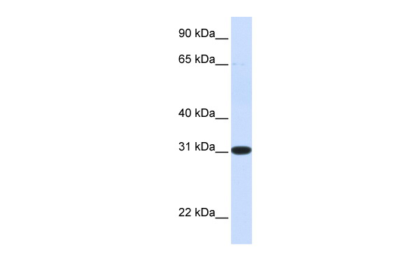
Human Liver; WB Suggested Anti-PBLD Antibody Titration: 0.2-1 ug/ml. ELISA Titer: 1:312500. Positive Control: Human Liver; PBLD antibody - N-terminal region (ARP56239_P050) in Human Liver cells using Western Blot

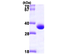
Recombinant human PBLD, 1-288 aa, His-tagged
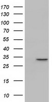
HEK293T cells were transfected with the pCMV6-ENTRY control (Left lane) or pCMV6-ENTRY PBLD (RC202328, Right lane) cDNA for 48 hrs and lysed. Equivalent amounts of cell lysates (5 ug per lane) were separated by SDS-PAGE and immunoblotted with anti-PBLD.

HEK293T cells transfected with either RC202328 overexpress plasmid(Red) or empty vector control plasmid(Blue) were immunostained by anti-PBLD antibody(TA501824), and then analyzed by flow cytometry.
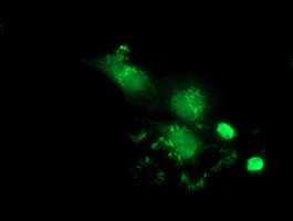
Anti-PBLD mouse monoclonal antibody (TA501840) immunofluorescent staining of COS7 cells transiently transfected by pCMV6-ENTRY PBLD(RC202328).
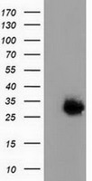
HEK293T cells were transfected with the pCMV6-ENTRY control (Left lane) or pCMV6-ENTRY PBLD (RC202328, Right lane) cDNA for 48 hrs and lysed. Equivalent amounts of cell lysates (5 ug per lane) were separated by SDS-PAGE and immunoblotted with anti-PBLD.
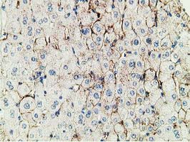
Immunohistochemical staining of paraffin-embedded Human liver tissue within the normal limits using anti-PBLD mouse monoclonal antibody. (TA501866)
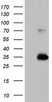
HEK293T cells were transfected with the pCMV6-ENTRY control (Left lane) or pCMV6-ENTRY PBLD (RC202328, Right lane) cDNA for 48 hrs and lysed. Equivalent amounts of cell lysates (5 ug per lane) were separated by SDS-PAGE and immunoblotted with anti-PBLD.
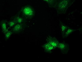
Anti-PBLD mouse monoclonal antibody (TA501994) immunofluorescent staining of COS7 cells transiently transfected by pCMV6-ENTRY PBLD(RC202328).
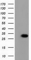
HEK293T cells were transfected with the pCMV6-ENTRY control (Left lane) or pCMV6-ENTRY PBLD (RC202328, Right lane) cDNA for 48 hrs and lysed. Equivalent amounts of cell lysates (5 ug per lane) were separated by SDS-PAGE and immunoblotted with anti-PBLD.
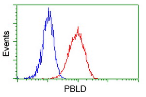
Flow cytometric Analysis of Hela cells, using anti-PBLD antibody(TA501994),(Red), compared to a nonspecific negative control antibody(TA50011),(Blue).
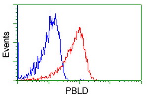
Flow cytometric Analysis of Jurkat cells, using anti-PBLD antibody(TA501994),(Red), compared to a nonspecific negative control antibody(TA50011),(Blue).
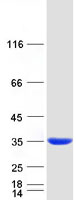
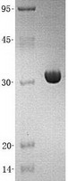
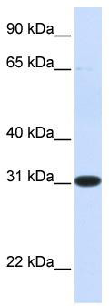
WB Suggested Anti-PBLD Antibody Titration: 0.2-1 ug/ml; ELISA Titer: 1:312500; Positive Control: Human Liver
Proteins & Growth Factors
| Catalog No. | Species | Pres. | Purity | Source | |
|---|---|---|---|---|---|
| TP720507 | MAWBP (transcript variant 1) |
||||
| Recombinant protein of human phenazine biosynthesis-like protein domain containing (PBLD), transcript variant 1 | |||||
 |
Human | > 95 % Preparation: . Purity Detail: >95% as determined by SDS-PAGE and Coomassie blue staining. |
E. coli |
10 µg /
€299.00
|
|
| OriGene Technologies, Inc. | |||||