PDCD6 antibody
Principal name
PDCD6 antibody
Alternative names for PDCD6 antibody
ALG2, ALG-2, Programmed cell death protein 6, Apoptosis-linked gene 2 protein, Probable calcium-binding protein ALG-2
Ncbi ID
Available reactivities
Available hosts
Available applications
Enzyme Immunoassay (E), Immunocytochemistry/Immunofluorescence (ICC/IF), Western blot / Immunoblot (WB), Paraffin Sections (P)
Background of PDCD6 antibody
- Kit Component:
- - KN312998G1, Pdcd6 gRNA vector 1 in pCas-Guide vector
- KN312998G2, Pdcd6 gRNA vector 2 in pCas-Guide vector
- KN312998D, donor vector containing left and right homologous arms and Luciferase-Puro functional cassette
- GE100003, scramble sequence in pCas-Guide vector - CRISPR Manual:
- CRISPR/CAS9 Genome Editing Manual
General readings
Okumura M., et al. (2009) Biochem. Biophys. Res. Commun. 386:237-241.
Suzuki H., et al. (2008), Structure 16:1562-1573.

Immunofluorescence of monoclonal antibody to PDCD6 on HeLa cell. [antibody concentration 10 ug/ml]

PDCD6 monoclonal antibody (M01), clone 2B4 Western Blot analysis of PDCD6 expression in HeLa ( Cat # L013V1 ).
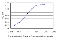
Detection limit for recombinant GST tagged PDCD6 is approximately 0.03ng/ml as a capture antibody.

Human lung carcinoma stained with Anti-PDCD6
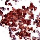
Immunohistochemical staining of PDCD6 on formalin fixed, paraffin embedded human lung cancer with PDCD6 polyclonal antibody.
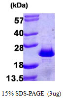
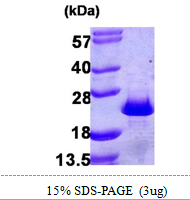
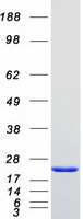
Recombinant human PDCD6, 1-191aa, His-tagged
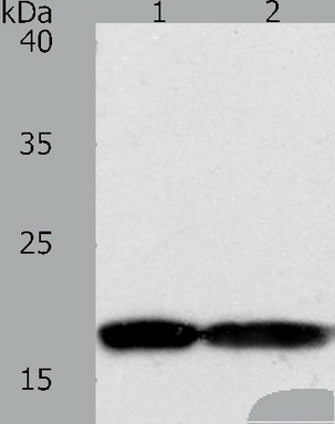
Predicted band size: 22kDa. Positive control: Jurkat and Hela cell lysate. Recommended dilution: 1/200-1000. (Gel: 10+12%SDS-PAGE Lane1: Jurkat cell lysate Lane2: JHela cell lysate Lysates: 50 ug per lane Primary antibody: 1/200 dilution Secondary antibody: Goat anti Rabbit IgG - H&L (HRP) at 1/8000 dilution Exposure time: 1 minute)
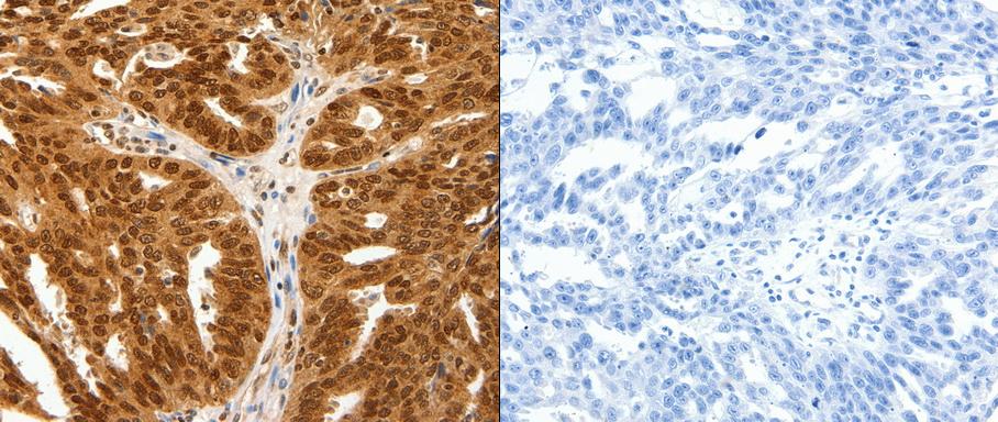
Predicted cell location: Cytoplasm, Nucleus. Positive control: Human ovarian cancer tissue. Recommended dilution: 1/25-100 The image on the left is immunohistochemistry of paraffin-embedded human ovarian cancer tissue using PDCD6 antibody at dilution 1/25, on the right is treated with the fusion protein. (Original magnification: x200)

Predicted band size: 22kDa. Positive control: Jurkat and hela cell, human fetal lung tissue and A549 cell lysate. Recommended dilution: 1/200-1000. (Gel: 10+12%SDS-PAGE Lane1-4:Jurkat and hela cell, human fetal lung tissue and A549 cell lysate Lysates: 50 ug per lane Primary antibody: 1/200 dilution Secondary antibody: Goat anti Rabbit IgG - H&L (HRP) at 1/8000 dilution Exposure time: 1 minute)
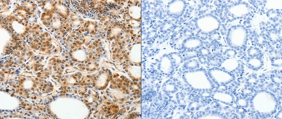
Predicted cell location: Cytoplasm, Nucleus. Positive control: Human thyroid cancer tissue. Recommended dilution: 1/25-100 The image on the left is immunohistochemistry of paraffin-embedded human thyroid cancer tissue using PDCD6 antibody at dilution 1/30, on the right is treated with the fusion protein. (Original magnification:x200)
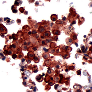
Human Lung Carcinoma stained with anti-PDCD6 antibody.
Primary Antibodies
| Catalog No. | Host | Iso. | Clone | Pres. | React. | Applications | |
|---|---|---|---|---|---|---|---|
| TA324255 | PDCD6 antibody |
||||||
| Anti-PDCD6 rabbit polyclonal antibody | |||||||
 |
Rabbit | IgG | Purified | Hu, Ms, Rt | E, P, WB |
0.1 ml /
€335.00
|
|
| OriGene Technologies, Inc. | |||||||
 +1 additional image +1 additional image |
|||||||
| TA324256 | PDCD6 antibody |
||||||
| Anti-PDCD6 rabbit polyclonal antibody | |||||||
 |
Rabbit | IgG | Purified | Hu, Ms, Rt | E, P, WB |
0.1 ml /
€335.00
|
|
| OriGene Technologies, Inc. | |||||||
| +1 additional image |
|||||||
Proteins & Growth Factors
| Catalog No. | Species | Pres. | Purity | Source | |
|---|---|---|---|---|---|
| TP302030 | PDCD6 |
||||
| Recombinant protein of human programmed cell death 6 (PDCD6) | |||||
 |
Human | > 80 % Preparation: Recombint protein was captured through anti-DDK affinity column followed by conventiol chromatography steps. Purity Detail: > 80% as determined by SDS-PAGE and Coomassie blue staining. |
HEK293 cells |
20 µg /
€748.00
|
|
| OriGene Technologies, Inc. | |||||