Peripherin antibody
Principal name
Peripherin antibody
Alternative names for Peripherin antibody
PRPH, NEF4, PRPH1
Ncbi ID
9606, NP_006253.2, 10116, NP_006253, 10090, NM_001163589, NM_013639, NM_001163588, NM_006262
Available reactivities
Available hosts
Available applications
Enzyme Immunoassay (E), Western blot / Immunoblot (WB), Paraffin Sections (P), ELISA (detection) (E(detection)), Frozen Sections (C), Immunocytochemistry/Immunofluorescence (ICC/IF)
Background of Peripherin antibody
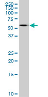
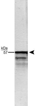
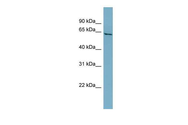
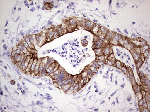
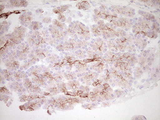
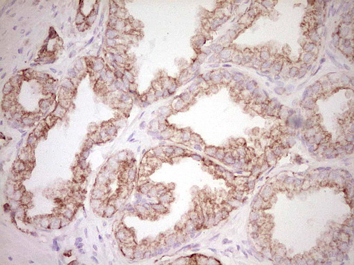
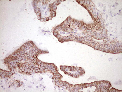
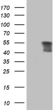
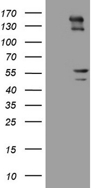
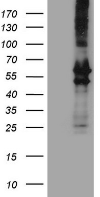
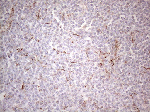
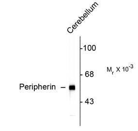
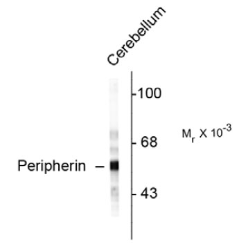
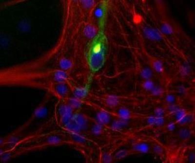
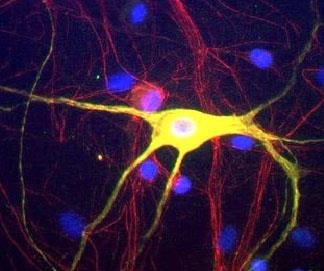
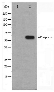
Peripherin is a ~57kDa intermediate filament subunit found initially in sensory neurons of the peripheral nervous systems, which gives the protein its name. Subsequently, peripherin was found in some sensory and other neurons of the central nervous system and also in PC12 cells. Peripherin is also expressed in certain neuroendocrine tumors and in the insulin producing cells of the pancreas. Peripherin belongs to the Class III family of intermediate filament subunits which also includes vimentin, glial fibrillary acidic protein (GFAP) and desmin. In contrast to the neurofilaments, peripherin is strongly up-regulated after nerve injury (1). Antibodies to peripherin can be used in identifying, classifying, and studying neurons throughout the nervous system. Peripherin is also a good diagnostic marker for ballooned axons seen in Lou Gehrig's disease (Amyotrophic lateral sclerosis) and some neuronally derived tumors (2). Autoantibodies to peripherin are frequently seen in the sera of patients with diabetes (3). Peripherin is not related to peripherin/RDS, a protein of the photoreceptor outer membrane mutations of which are causative of certain forms of slow retinal degeneration.

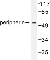
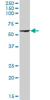
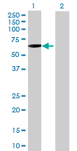
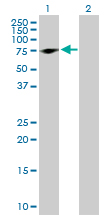

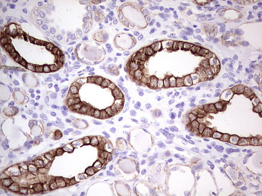
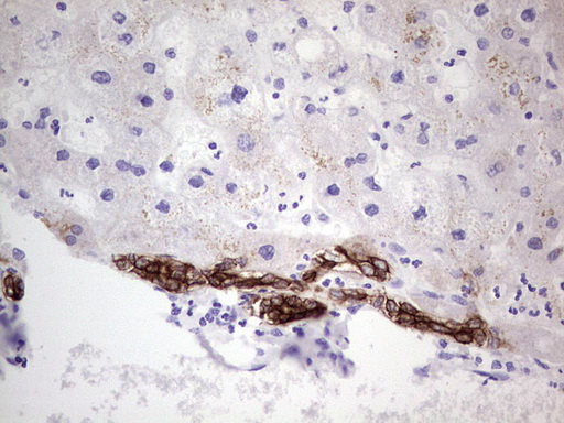
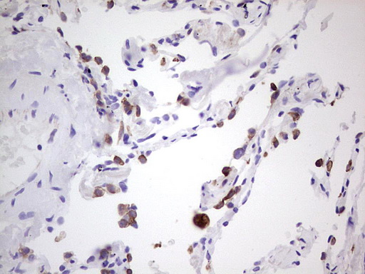
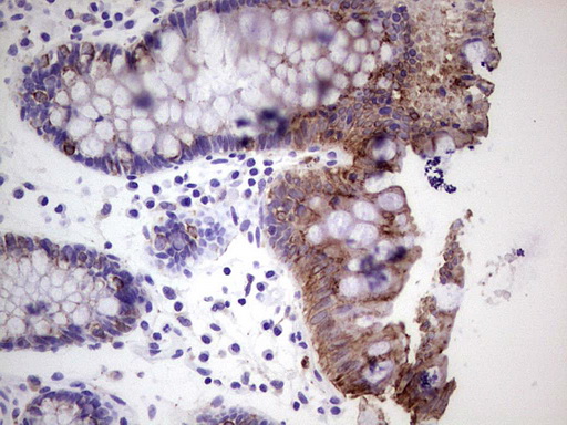

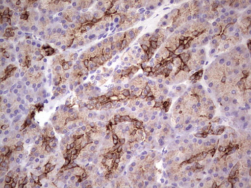
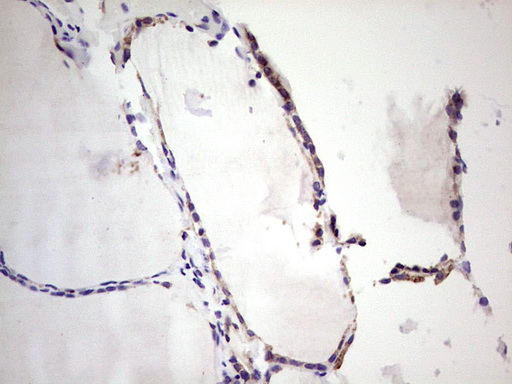
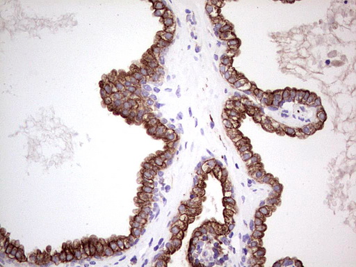
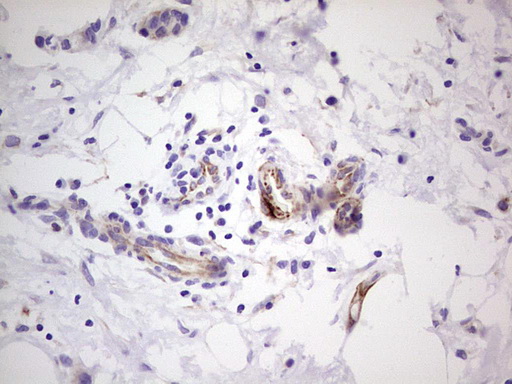


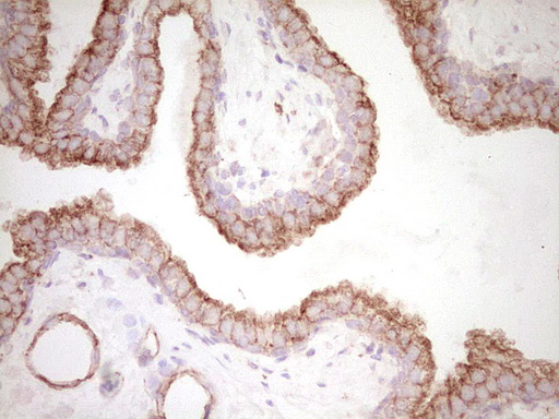
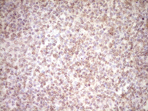
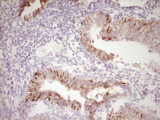

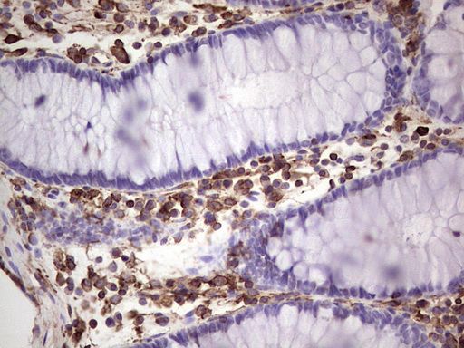
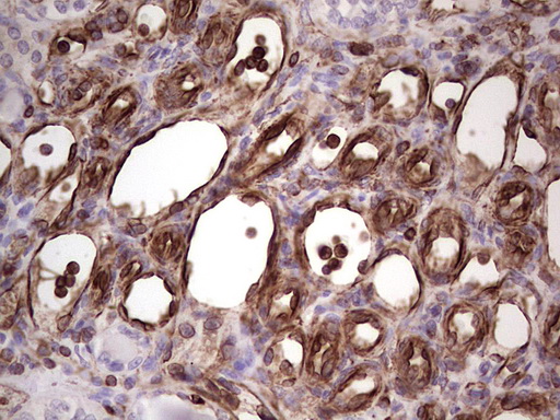
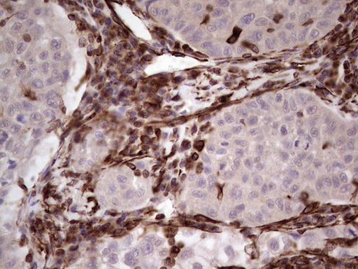
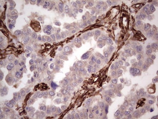
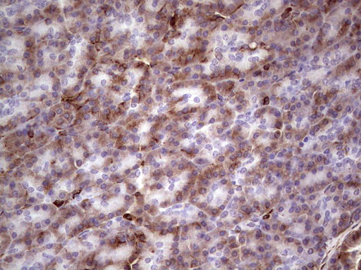

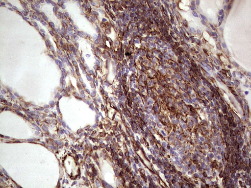
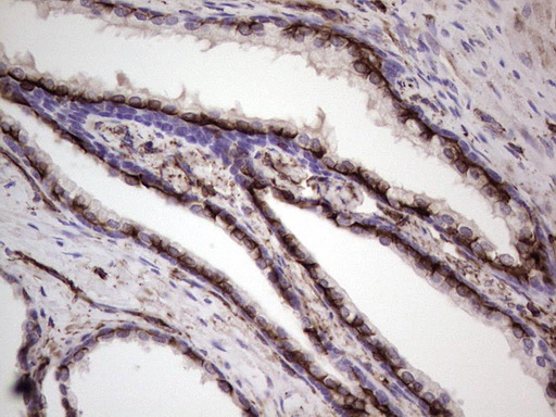
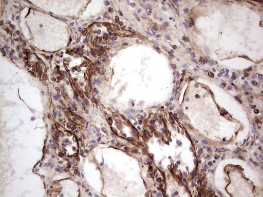
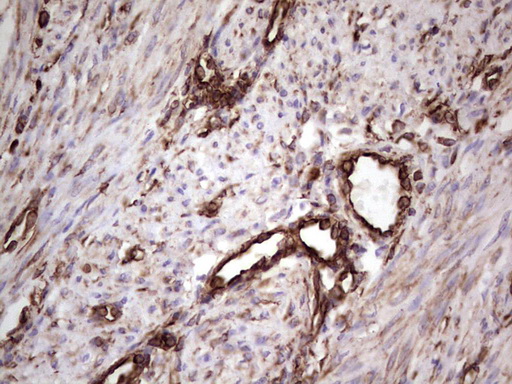
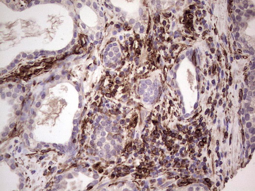
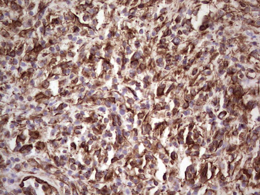

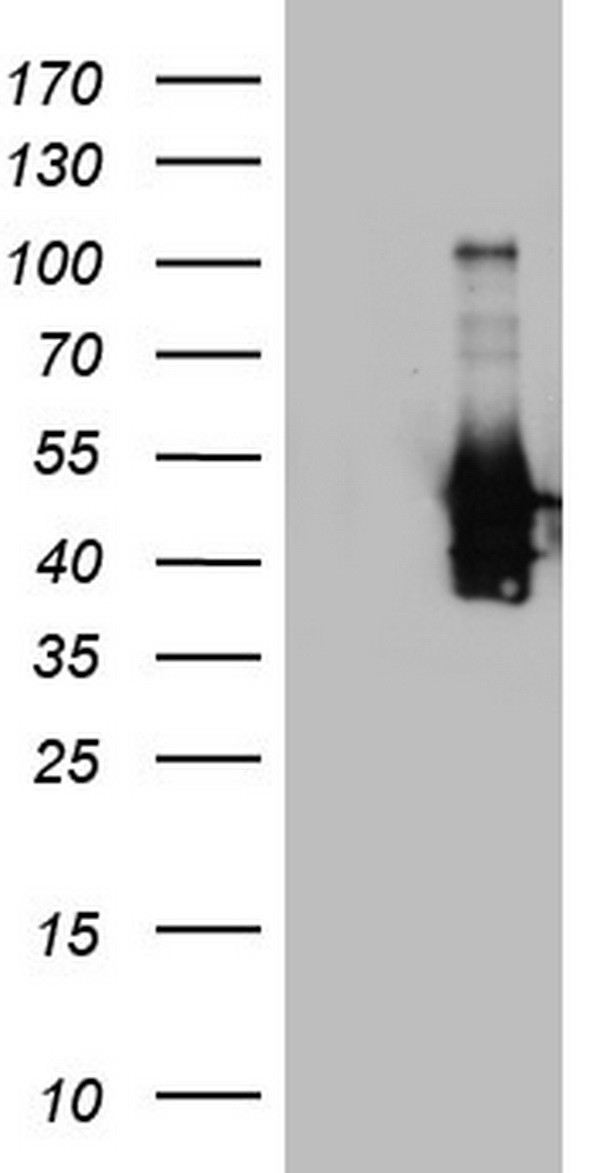
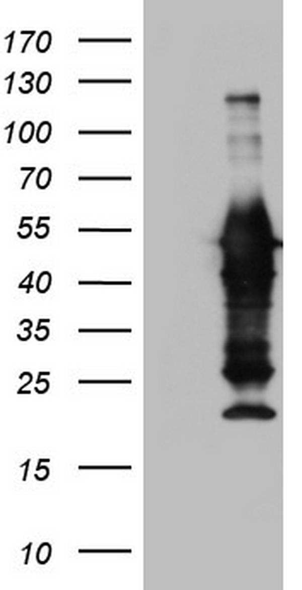

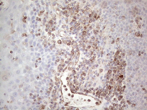
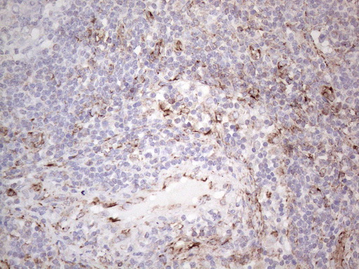
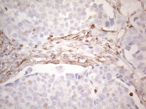
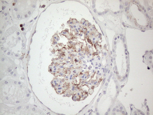
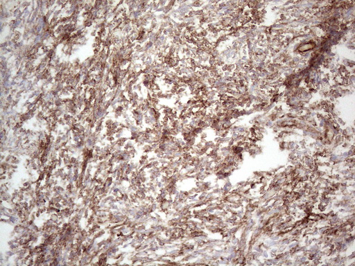
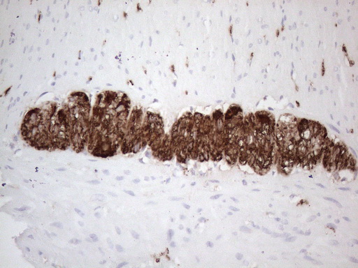
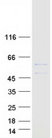
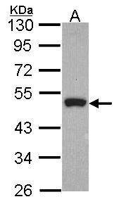


- Quick-Filters:
- Highly Validated (5)
Primary Antibodies
| Catalog No. | Host | Iso. | Clone | Pres. | React. | Applications | |
|---|---|---|---|---|---|---|---|
| UM800094 | Peripherin antibody |
||||||
| PRPH mouse monoclonal antibody,clone UMAB202 | |||||||
 |
Mouse | IgG1 | UMAB202 | Purified | Hu | P |
0.1 ml /
€468.00
|
| OriGene Technologies, Inc. | |||||||
 +6 additional images +6 additional images |
Highly validated antibody!
|
||||||
| UM870094 | Peripherin antibody |
||||||
| PRPH mouse monoclonal antibody,clone UMAB202 | |||||||
|
Mouse | IgG1 | UMAB202 | Purified | Hu | P |
30 µl /
€180.00
|
| OriGene Technologies, Inc. | |||||||
| +6 additional images |
Highly validated antibody!
|
||||||
| TA806181 | Peripherin antibody |
||||||
| PRPH mouse monoclonal antibody, clone OTI3F4 (formerly 3F4) | |||||||
 |
Mouse | IgG1 | OTI3F4 | Purified | Hu | P |
0.1 ml /
€360.00
|
| OriGene Technologies, Inc. | |||||||
| +9 additional images |
Highly validated antibody!
|
||||||
| TA806182 | Peripherin antibody |
||||||
| PRPH mouse monoclonal antibody, clone OTI2D5 (formerly 2D5) | |||||||
 |
Mouse | IgG1 | OTI2D5 | Purified | Hu | P, WB |
0.1 ml /
€360.00
|
| OriGene Technologies, Inc. | |||||||
| +8 additional images |
Highly validated antibody!
|
||||||
| TA806183 | Peripherin antibody |
||||||
| PRPH mouse monoclonal antibody, clone OTI1E10 (formerly 1E10) | |||||||
 |
Mouse | IgG1 | OTI1E10 | Purified | Hu | P, WB |
0.1 ml /
€360.00
|
| OriGene Technologies, Inc. | |||||||
| +13 additional images |
Highly validated antibody!
|
||||||
| TA309213 | Peripherin antibody |
||||||
| Mouse Anti-Peripherin ms Antibody | |||||||
 |
Mouse | IgG1 | 7C5 | Supernatant | Mam | C, ICC/IF, WB |
0.1 ml /
€405.00
|
| OriGene Technologies, Inc. | |||||||
| TA336620 | Peripherin antibody |
||||||
| Mouse Monoclonal Peripherin Antibody (7C5) | |||||||
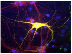
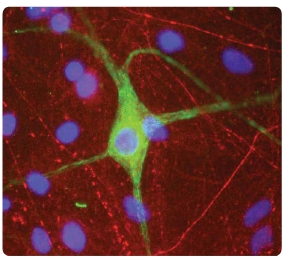
![Immunocytochemistry/Immunofluorescence: Peripherin Antibody (7C5) [NB300-220] - Mixed neuron/glia cultures from newborn rat brain stained with NB300-220 antibody to peripherin (green) and rabbit polyclonal antibody to NF-L NBP1-05218 (red channel). A clas](https://m1.acris-antibodies.com/catalog/product/cache/1/small_image/50x50/9df78eab33525d08d6e5fb8d27136e95/OriGene/NB300-220-0.2ml-1-F.jpg) |
Mouse | IgG1 | 7C5 | Purified | Chk, Hu, Mam, Ms, Rt | ICC/IF, WB |
0.5 ml /
€220.00
|
| OriGene Technologies, Inc. | |||||||
| TA806185 | Peripherin antibody |
||||||
| PRPH mouse monoclonal antibody, clone OTI3H1 (formerly 3H1) | |||||||
 |
Mouse | IgG1 | OTI3H1 | Purified | Hu | WB |
0.1 ml /
€360.00
|
| OriGene Technologies, Inc. | |||||||
|
|||||||
| TA806223 | Peripherin antibody |
||||||
| PRPH mouse monoclonal antibody, clone OTI7D8 (formerly 7D8) | |||||||
 |
Mouse | IgG1 | OTI7D8 | Purified | Hu | WB |
0.1 ml /
€360.00
|
| OriGene Technologies, Inc. | |||||||
|
|||||||
| TA806224 | Peripherin antibody |
||||||
| PRPH mouse monoclonal antibody, clone OTI4A2 (formerly 4A2) | |||||||
 |
Mouse | IgG1 | OTI4A2 | Purified | Hu | WB |
0.1 ml /
€360.00
|
| OriGene Technologies, Inc. | |||||||
|
|||||||
| TA806226 | Peripherin antibody |
||||||
| PRPH mouse monoclonal antibody, clone OTI7E4 (formerly 7E4) | |||||||
 |
Mouse | IgG2b | OTI7E4 | Purified | Hu | WB |
0.1 ml /
€360.00
|
| OriGene Technologies, Inc. | |||||||
|
|||||||
| TA806240 | Peripherin antibody |
||||||
| PRPH mouse monoclonal antibody, clone OTI8G3 (formerly 8G3) | |||||||
 |
Mouse | IgG2a | OTI8G3 | Purified | Hu | WB |
0.1 ml /
€360.00
|
| OriGene Technologies, Inc. | |||||||
|
|||||||