PPWD1 antibody
Principal name
PPWD1 antibody
Alternative names for PPWD1 antibody
KIAA0073, Spliceosome-associated cyclophilin, Peptidylprolyl isomerase domain and WD repeat-containing protein 1
Ncbi ID
9606, NP_056157, NP_056157.1, NM_015342, NP_766395.2, NM_172807
Available reactivities
Available hosts
Available applications
ELISA (detection) (E(detection)), Western blot / Immunoblot (WB), Immunocytochemistry/Immunofluorescence (ICC/IF), Paraffin Sections (P), Flow Cytometry (F)
Background of PPWD1 antibody
- Kit Component:
- - KN313800G1, Ppwd1 gRNA vector 1 in pCas-Guide vector
- KN313800G2, Ppwd1 gRNA vector 2 in pCas-Guide vector
- KN313800D, donor vector containing left and right homologous arms and Luciferase-Puro functional cassette
- GE100003, scramble sequence in pCas-Guide vector - CRISPR Manual:
- CRISPR/CAS9 Genome Editing Manual
General readings
Jurica,M.S., (2002) RNA 8 (4), 426-439
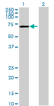
Western Blot analysis of PPWD1 expression in transfected 293T cell line by PPWD1 MaxPab polyclonal antibody.Lane 1: PPWD1 transfected lysate(71.06 KDa).Lane 2: Non-transfected lysate.

Human 293T; WB Suggested Anti-PPWD1 Antibody Titration: 0.2-1 ug/ml. ELISA Titer: 1:312500. Positive Control: 293T cell lysate. PPWD1 is supported by BioGPS gene expression data to be expressed in HEK293T.; PPWD1 antibody - middle region (ARP55238_P050) in Human 293T cells using Western Blot
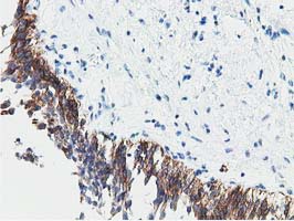
Immunohistochemical staining of paraffin-embedded Human bladder tissue within the normal limits using anti-PPWD1 mouse monoclonal antibody. (TA501891)

Immunohistochemical staining of paraffin-embedded Human Kidney tissue within the normal limits using anti-PPWD1 mouse monoclonal antibody. (TA501891)
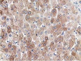
Immunohistochemical staining of paraffin-embedded Human liver tissue within the normal limits using anti-PPWD1 mouse monoclonal antibody. (TA501891)

Anti-PPWD1 mouse monoclonal antibody (TA501891) immunofluorescent staining of COS7 cells transiently transfected by pCMV6-ENTRY PPWD1(RC204570).

HEK293T cells were transfected with the pCMV6-ENTRY control (Left lane) or pCMV6-ENTRY PPWD1 (RC204570, Right lane) cDNA for 48 hrs and lysed. Equivalent amounts of cell lysates (5 ug per lane) were separated by SDS-PAGE and immunoblotted with anti-PPWD1.
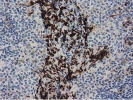
Immunohistochemical staining of paraffin-embedded Human lymph node tissue within the normal limits using anti-PPWD1 mouse monoclonal antibody. (TA501947)
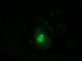
Anti-PPWD1 mouse monoclonal antibody (TA501947) immunofluorescent staining of COS7 cells transiently transfected by pCMV6-ENTRY PPWD1(RC204570).
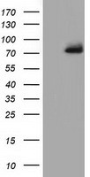
HEK293T cells were transfected with the pCMV6-ENTRY control (Left lane) or pCMV6-ENTRY PPWD1 (RC204570, Right lane) cDNA for 48 hrs and lysed. Equivalent amounts of cell lysates (5 ug per lane) were separated by SDS-PAGE and immunoblotted with anti-PPWD1.
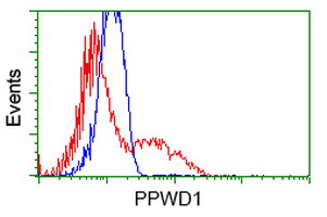
HEK293T cells transfected with either RC204570 overexpress plasmid(Red) or empty vector control plasmid(Blue) were immunostained by anti-PPWD1 antibody(TA501947), and then analyzed by flow cytometry.
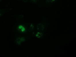
Anti-PPWD1 mouse monoclonal antibody (TA501948) immunofluorescent staining of COS7 cells transiently transfected by pCMV6-ENTRY PPWD1(RC204570).
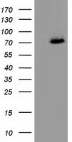
HEK293T cells were transfected with the pCMV6-ENTRY control (Left lane) or pCMV6-ENTRY PPWD1 (RC204570, Right lane) cDNA for 48 hrs and lysed. Equivalent amounts of cell lysates (5 ug per lane) were separated by SDS-PAGE and immunoblotted with anti-PPWD1.

HEK293T cells transfected with either RC204570 overexpress plasmid(Red) or empty vector control plasmid(Blue) were immunostained by anti-PPWD1 antibody(TA501948), and then analyzed by flow cytometry.
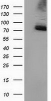
HEK293T cells were transfected with the pCMV6-ENTRY control (Left lane) or pCMV6-ENTRY PPWD1 (RC204570, Right lane) cDNA for 48 hrs and lysed. Equivalent amounts of cell lysates (5 ug per lane) were separated by SDS-PAGE and immunoblotted with anti-PPWD1.
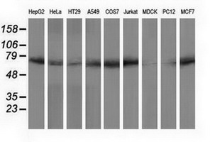
Western blot analysis of extracts (35ug) from 9 different cell lines by using anti-PPWD1 monoclonal antibody.

HEK293T cells were transfected with the pCMV6-ENTRY control (Left lane) or pCMV6-ENTRY PPWD1 (RC204570, Right lane) cDNA for 48 hrs and lysed. Equivalent amounts of cell lysates (5 ug per lane) were separated by SDS-PAGE and immunoblotted with anti-PPWD1.
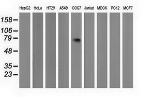
Western blot analysis of extracts (35ug) from 9 different cell lines by using anti-PPWD1 monoclonal antibody.


WB Suggested Anti-PPWD1 Antibody Titration: 0.2-1 ug/ml; ELISA Titer: 1: 312500; Positive Control: 293T cell lysatePPWD1 is supported by BioGPS gene expression data to be expressed in HEK293T
- Quick-Filters:
- Highly Validated (2)
Primary Antibodies
| Catalog No. | Host | Iso. | Clone | Pres. | React. | Applications | |
|---|---|---|---|---|---|---|---|
| TA501891 | PPWD1 antibody |
||||||
| PPWD1 mouse monoclonal antibody, clone OTI1B2 (formerly 1B2) | |||||||
 |
Mouse | IgG1 | OTI1B2 | Purified | Hu | ICC/IF, P, WB |
0.1 ml /
€360.00
|
| OriGene Technologies, Inc. | |||||||
 +4 additional images +4 additional images |
Highly validated antibody!
|
||||||
| TA501947 | PPWD1 antibody |
||||||
| PPWD1 mouse monoclonal antibody, clone OTI1B8 (formerly 1B8) | |||||||
 |
Mouse | IgG1 | OTI1B8 | Purified | Hu | F, ICC/IF, P, WB |
0.1 ml /
€360.00
|
| OriGene Technologies, Inc. | |||||||
| +3 additional images |
Highly validated antibody!
|
||||||
| TA501948 | PPWD1 antibody |
||||||
| PPWD1 mouse monoclonal antibody, clone OTI1G6 (formerly 1G6) | |||||||
 |
Mouse | IgG1 | OTI1G6 | Purified | Hu | F, ICC/IF, WB |
0.1 ml /
€360.00
|
| OriGene Technologies, Inc. | |||||||
| +2 additional images |
|
||||||
| TA502069 | PPWD1 antibody |
||||||
| PPWD1 mouse monoclonal antibody, clone OTI1G5 (formerly 1G5) | |||||||
 |
Mouse | IgG2b | OTI1G5 | Purified | Can, Hu, Mky, Rt | WB |
0.1 ml /
€379.00
|
| OriGene Technologies, Inc. | |||||||
| +1 additional image |
|
||||||
| TA502076 | PPWD1 antibody |
||||||
| PPWD1 mouse monoclonal antibody, clone OTI2B1 (formerly 2B1) | |||||||
 |
Mouse | IgG1 | OTI2B1 | Purified | Hu, Mky | WB |
0.1 ml /
€360.00
|
| OriGene Technologies, Inc. | |||||||
| +1 additional image |
|
||||||

Proteins & Growth Factors
| Catalog No. | Species | Pres. | Purity | Source | |
|---|---|---|---|---|---|
| TP304570 | PPWD1 |
||||
| Recombinant protein of human peptidylprolyl isomerase domain and WD repeat containing 1 (PPWD1) | |||||
 |
Human | > 80 % Preparation: Recombint protein was captured through anti-DDK affinity column followed by conventiol chromatography steps. Purity Detail: > 80% as determined by SDS-PAGE and Coomassie blue staining. |
HEK293 cells |
20 µg /
€748.00
|
|
| OriGene Technologies, Inc. | |||||
Lysates
| Catalog No. | ||
|---|---|---|
| LY414604 | PPWD1 overexpression lysate |
|
 |
0.1 mg /
€295.00
|
|
| OriGene Technologies, Inc. |
Mouse Lenti ORF Particles
| Catalog No. | ||
|---|---|---|
| MR215653L2V | Lenti ORF particles, Ppwd1 (GFP-tagged) - Mouse peptidylprolyl isomerase domain and WD repeat containing 1 (Ppwd1), 200 uL, >10^7 TU/mL |
|
|
200 µl /
€1,400.00
|
|
| OriGene Technologies, Inc. | ||
| MR215653L1V | Lenti ORF particles, Ppwd1 (Myc-DDK-tagged) - Mouse peptidylprolyl isomerase domain and WD repeat containing 1 (Ppwd1), 200 uL, >10^7 TU/mL |
|
|
200 µl /
€1,250.00
|
|
| OriGene Technologies, Inc. | ||