PROX-1 antibody
Principal name
PROX-1 antibody
Alternative names for PROX-1 antibody
PROX1, Prospero homeobox protein 1, Homeobox prospero-like protein PROX1
Ncbi ID
NP_002754.2, 9606, NP_002754, NM_002763, NP_032963.1, NM_008937
Available reactivities
Available hosts
Available applications
Frozen Sections (C), Western blot / Immunoblot (WB), Paraffin Sections (P), Immunocytochemistry/Immunofluorescence (ICC/IF), Enzyme Immunoassay (E)
Background of PROX-1 antibody
The expression pattern of the Prox1 homeo box gene suggests that it has a role in a variety of embryonic tissues, including lens. Analysis of mRNA reveals that Prox mRNA is present in many different human tissues and that lens demonstrated the highest level. Homozygous Prox1-null mice die at midgestation from multiple developmental defects, and a targeted effect on lens development has been reported. Prox1 inactivation caused abnormal cellular proliferation, downregulated expression of the cell cycle inhibitors Cdkn1b and Cdkn1c, misexpression of E-cadherin, and excessive apoptosis. Consequently, mutant lens cells failed to polarize and elongate properly, resulting in a hollow lens. The Prox1 gene is expressed in a subpopulation of endothelial cells that by budding and sprouting give rise to the lymphatic system. Prox1 appears to be a specific and required regulator of the development of the lymphatic system. Prox1 also has been document to be required for hepatocyte migration in the mouse. Loss of Prox1 results in a smaller liver with a reduced population of clustered hepatocytes. The homeodomain protein Prox1 regulates the egress of progenitor cells from the cell cycle in the embryonic mouse retina. Cells lacking Prox1 are less likely to stop dividing, and ectopic expression of Prox1 forces progenitor cells to exit the cell cycle. Prox1 acts as a key participant in progenitor-cell proliferation and cell-fate determination in the vertebrate retina.
General readings
1. Joshua D. Webster, Margaret A. Miller, Dee DuSold, and José Ramos-Vara. Effects of Prolonged Formalin-fixation on Diagnostic Immunohistochemistry in Domestic Animals.
J. Histochem. Cytochem., Apr 2009; 10.1369/jhc.2009.953877.
2. Ohad Shaham, April N. Smith, Michael L. Robinson, Makoto M. Taketo, Richard A. Lang, and Ruth Ashery-Padan. Pax6 is essential for lens fiber cell differentiation. Development, Aug 2009; 136: 2567-2578.
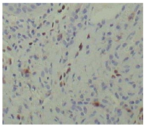
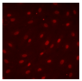
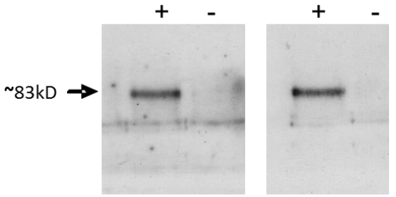
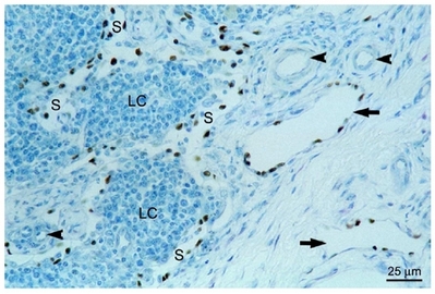
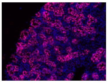
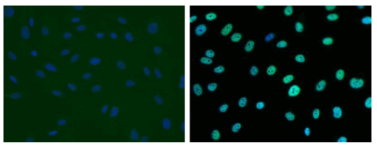
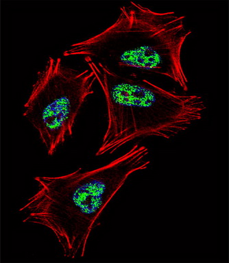
Both experiments were performed by Andreia Margarido (de Santa Barbara’s team); INSERM U1046, Montpellier, France
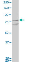

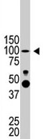
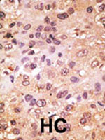
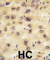

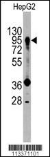
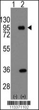
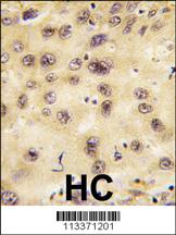


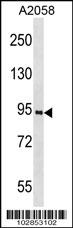


- Quick-Filters:
- Cited (2)
- Highly Validated (1)
Primary Antibodies
| Catalog No. | Host | Iso. | Clone | Pres. | React. | Applications | |
|---|---|---|---|---|---|---|---|
| DP3501 | PROX-1 (C-term) antibody |
||||||
 |
Rabbit | Serum | Hu, Ms, Rt, Ze | C, P, WB |
0.2 ml /
€400.00
|
||
| OriGene Technologies GmbH | |||||||
 +1 additional image +1 additional image |
Published recently! |
||||||
| DP3516 | PROX-1 (C-term) antibody |
||||||
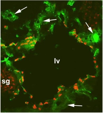
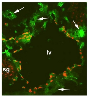
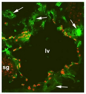
![Immunofluorescence with a Frozen Section of a Mouse embryo at day 13.5 with anti-CD31 Antibody [Green] and anti-Human Prox-1 Antibody [red] (Cat.-No DP3516). It shows a large lymphatic vessel (lv), blood vessels (arrows) and a sympathetic ganglion (sg) which is also positiv for Prox-1.
The experiment was performed by PD. Dr. Jörg Wilting, Pädiatrie I, Zentrum für Kinderheilkunde und Jugendmedizin in Göttingen, Germany.](https://m1.acris-antibodies.com/catalog/product/cache/1/small_image/50x50/9df78eab33525d08d6e5fb8d27136e95/0092801.jpeg) |
Rabbit | Purified | Hu, Ms, Rt | ICC/IF, P, WB |
0.2 mg /
€350.00
|
||
| OriGene Technologies GmbH | |||||||
| +1 additional image |
Published recently! |
||||||
| DP3516P | PROX-1 (C-term) antibody |
||||||
 |
Rabbit | IgG | Aff - Purified | Hu, Ms, Rt | C, E, ICC/IF, P, WB |
50 µg /
€280.00
|
|
| OriGene Technologies GmbH | |||||||
| +6 additional images |
Highly validated antibody! |
||||||
| TA302031 | PROX-1 antibody |
||||||
| Rabbit Polyclonal Antibody against PROX1 (Center) | |||||||
 |
Rabbit | Ig | Aff - Purified | Hu, Ms | P, WB |
0.4 ml /
€415.00
|
|
| OriGene Technologies, Inc. | |||||||
| +2 additional images |
|||||||
| TA309222 | PROX-1 antibody |
||||||
| mouse Anti-Prox1 Antibody | |||||||
 |
Mouse | IgG | 5G10 | Aff - Purified | Chk, Hu, Ms, Rt | P |
0.1 ml /
€405.00
|
| OriGene Technologies, Inc. | |||||||
| TA324702 | PROX-1 (C-term) antibody |
||||||
| Rabbit polyclonal PROX1 Antibody (C-term) | |||||||
 |
Rabbit | Ig | Purified | Hu, Ms | P, WB |
0.4 ml /
€415.00
|
|
| OriGene Technologies, Inc. | |||||||
| +2 additional images |
|||||||
| TA351557 | PROX-1 antibody |
||||||
| Rabbit Polyclonal Anti-PROX1 Antibody | |||||||
 |
Rabbit | IgG | Purified | Hu, Ms | E, P, WB |
0.1 ml /
€335.00
|
|
| OriGene Technologies, Inc. | |||||||
| +1 additional image |
|||||||
| AP11899PU-N | PROX-1 (N-term) antibody |
||||||
 |
Rabbit | Ig | Purified | Hu, Ms | WB |
0.4 ml /
€415.00
|
|
| OriGene Technologies GmbH | |||||||
| AP26049PU-N | PROX-1 (723-727) antibody |
||||||
 |
Rabbit | Aff - Purified | Hu, Ms | WB |
0.1 mg /
€300.00
|
||
| OriGene Technologies GmbH | |||||||
| AP26049PU-S | PROX-1 (723-727) antibody |
||||||
|
Rabbit | Aff - Purified | Hu, Ms | WB |
50 µg /
€230.00
|
||
| OriGene Technologies GmbH | |||||||
| DP3501PS | PROX-1 (C-term) antibody |
||||||
 |
Rabbit | Purified | Fi, Hu, Ms, Rt | C, P, WB |
0.1 mg /
€400.00
|
||
| OriGene Technologies GmbH | |||||||
| DP3516B | PROX-1 (C-term) antibody |
||||||
|
Rabbit | Biotin | Hu, Ms, Rt | ICC/IF |
50 µg /
€350.00
|
||
| OriGene Technologies GmbH | |||||||
| DP3516S | PROX-1 (C-term) antibody |
||||||
![Figure 1. Immunofluorescence with a Frozen Section of a Mouse embryo at day 13.5 with anti-CD31 [green] and anti-Human Prox-1 [red] (Cat.-No DP3516S). It shows a large lymphatic vessel (lv), blood vessels (arrows) and a sympathetic ganglion (sg) which is also positiv for Prox-1 (Fig.1). The experiment was performed by PD. Dr. Jörg Wilting, Pädiatrie I, Zentrum für Kinderheilkunde und Jugendmedizin in Göttingen, Germany.](https://m1.acris-antibodies.com/catalog/product/cache/1/small_image/50x50/9df78eab33525d08d6e5fb8d27136e95/0219346.jpeg) |
Rabbit | Purified | Hu, Ms, Rt | C, ICC/IF, WB |
0.1 mg /
€270.00
|
||
| OriGene Technologies GmbH | |||||||
Lysates
| Catalog No. | ||
|---|---|---|
| LY400978 | PROX1 overexpression lysate |
|
|
0.1 mg /
€295.00
|
|
| OriGene Technologies, Inc. |
Mouse Lenti ORF Particles
| Catalog No. | ||
|---|---|---|
| MR210370L2V | Lenti ORF particles, Prox1 (GFP-tagged) - Mouse prospero-related homeobox 1 (Prox1), 200 uL, >10^7 TU/mL |
|
|
200 µl /
€1,530.00
|
|
| OriGene Technologies, Inc. | ||
| MR210370L1V | Lenti ORF particles, Prox1 (Myc-DDK-tagged) - Mouse prospero-related homeobox 1 (Prox1), 200 uL, >10^7 TU/mL |
|
|
200 µl /
€1,370.00
|
|
| OriGene Technologies, Inc. | ||

