RFP-Tag antibody
Principal name
RFP-Tag antibody
Alternative names for RFP-Tag antibody
Red fluorescent protein Tag, DsRed Tag
SwissProt ID
Q9U6Y8 (Dissp)
Ncbi ID
Available reactivities
Available hosts
Available applications
Frozen Sections (C), Enzyme Immunoassay (E), Immunocytochemistry/Immunofluorescence (ICC/IF), Western blot / Immunoblot (WB), Immunoprecipitation (IP), Paraffin Sections (P), Flow Cytometry (F), Dot blot (Dot)
Background of RFP-Tag antibody
Epitope tagging is a powerful and versatile strategy for detecting and purifying proteins expressed by cloned genes. To utilize this feature, protein expression vectors are typically engineered with a nucleotide sequence that encodes the peptide epitope tag. The gene of interest is cloned in-frame relative to the tag and, upon expression, the protein of interest is synthesized as a fusion protein with the peptide tag. Fusion protein detection and/or purification is mediated by highly specific antibodies to the engineered peptide, thus eliminating the need for antibodies to proteins from each newly cloned gene. Commonly used epitope tags include glutathione-S-transferase (GST), c-myc, 6-histidine (6X-His), FLAG, green fluorescent protein (GFP), red fluorescent protein (RFP, DSRed), maltose binding protein (MBP), influenza A virus haemagglutinin (HA), b-galactosidase, and GAL4.
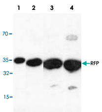

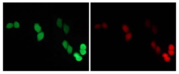
*This transfectant is provided by RIKEN.

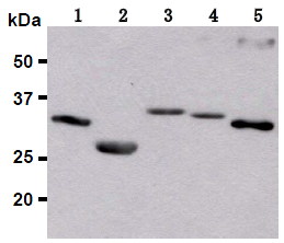

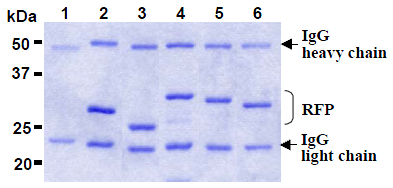
Immunoprecipitation of DsRed (1, 2), mRFP1* (3), mCherry* (4), mOrange* (5) and mPlum* (6) with isotype control (1) or AM26655AG-N (2-6). After immunoprecipitated with the antibody, immunocomplex was resolved on SDS-PAGE and stained with CBB. *Sample number (3) to (6) are provided by RIKEN.

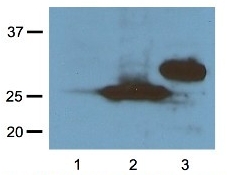
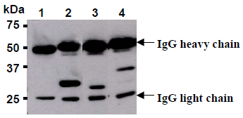
Immunoprecipitation of DsRed (1, 2), mRFP1* (3) and mCherry* (4) with isotype control (1) or AM26655AG-N (2-4). After immunoprecipitated with the antibody, immunocomplex was resolved on SDS-PAGE and immunoblotted with anti-RFP monoclonal antibody. *Sample number (3) and (4) are provided by RIKEN.
- Quick-Filters:
- Cited (1)
- Highly Validated (1)
Primary Antibodies
| Catalog No. | Host | Iso. | Clone | Pres. | React. | Applications | |
|---|---|---|---|---|---|---|---|
| AP09229PU-N | RFP-Tag (Ads. to Hu, Ms, Rt Serum Proteins) antibody |
||||||
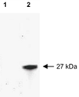
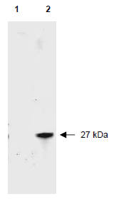
![Western blot of RFP recombinant protein detected with polyclonal anti-RFP antibody. Lane 1 shows no reaction against a GFP recombinant protein present in 10 μg of HeLa cell extract. Lane 2 shows a single band detected in 10 μg of a HeLa lysate containing RFP recombinant protein as a 27 kDa band. A 4-12% Bis-Tris gradient gel was used for SDS-PAGE. The membrane was blocked and then probed with Anti-RFP diluted 1:2,500 for 1 h at RT followed by washes and reaction with a 1:5,000 dilution of IRDye(TM)800 conjugated Goat-a-Rabbit IgG [H&L]. IRDye(TM)800 fluorescence image was captured using the Odyssey® Infrared Imaging System developed by LI-COR. IRDye is a trademark of LI-COR, Inc. Other detection systems will yield similar results.](https://m1.acris-antibodies.com/catalog/product/cache/1/small_image/50x50/9df78eab33525d08d6e5fb8d27136e95/0112253.png) |
Rabbit | IgG | Aff - Purified | E, P, WB |
0.1 mg /
€350.00
|
||
| OriGene Technologies GmbH | |||||||
 +4 additional images +4 additional images |
Published recently! Highly validated antibody! |
||||||
| AM20837PU-N | RFP-Tag antibody |
||||||
 |
Mouse | IgG1 | RFP. H8 | Aff - Purified | Dot, IP, WB |
0.1 mg /
€360.00
|
|
| OriGene Technologies GmbH | |||||||
| AM26650AF-N | RFP-Tag antibody |
||||||
 |
Mouse | IgG1 | 8D6 | Azide Free | ICC/IF, WB |
0.1 ml /
€430.00
|
|
| OriGene Technologies GmbH | |||||||
| +2 additional images |
|||||||
| AM26655AF-N | RFP-Tag antibody |
||||||
 |
Mouse | IgG2a | 3G5 | Azide Free | IP |
0.1 mg /
€340.00
|
|
| OriGene Technologies GmbH | |||||||
| +1 additional image |
|||||||
| AM26655AG-N | RFP-Tag antibody |
||||||
 |
Mouse | IgG2a | 3G5 | Agarose | IP |
0.2 ml /
€375.00
|
|
| OriGene Technologies GmbH | |||||||
| +1 additional image |
|||||||
| AP09229BT-N | RFP-Tag (Ads. to Hu, Ms, Rt Serum Proteins) antibody |
||||||
![Western blot of RFP recombinant protein detected with anti-RFP antibody. Lane 1 shows no reaction against a GFP recombinant protein present in 10 μg of HeLa cell extract. Lane 2 shows a single band detected in 10 μg of a HeLa lysate containing RFP recombinant protein. Separation was achieved using a 4-12% Bis Tris gradient gel followed by transfer to nitrocellulose and blocking.The membrane was probed with the primary antibody diluted 1:2,500 for 1 h at room temperature followed by washes and reaction with a 1:5,000 dilution of IRDye(TM)800 conjugated Goat-a-Rabbit IgG [H&L]. IRDye(TM)800 fluorescence image was captured using the Odyssey® Infrared Imaging System developed by LI-COR. IRDye is a trademark of LI-COR, Inc. Other detection systems will yield similar results.](https://m1.acris-antibodies.com/catalog/product/cache/1/small_image/50x50/9df78eab33525d08d6e5fb8d27136e95/0163852.png) |
Rabbit | IgG | Biotin | C, E, WB |
0.1 mg /
€515.00
|
||
| OriGene Technologies GmbH | |||||||
| AP09229FC-N | RFP-Tag (Ads. to Hu, Ms, Rt Serum Proteins) antibody |
||||||
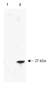
![Western blot of RFP recombinant protein detected withanti-RFP antibody. Lane 1 shows no reaction against a GFP recombinant protein present in 10 μg of HeLa cell extract. Lane 2 shows a single band detected in 10 μg of a HeLa lysate containing RFP recombinant protein. Anti-RFP detects a 27 kDa band corresponding to the epitope tag RFP. A 4-12% Bis-Tris gradient gel (Invitrogen) was used for SDSPAGE. The protein was transferred to nitrocellulose using standard methods. After blocking the membrane was probed with the primary antibody diluted 1:2,500 for 1 h at room temperature followed by washes and reaction with a 1:5,000 dilution of IRDye(TM)800 conjugated Goat-a-Rabbit IgG [H&L]. IRDye(TM)800 fluorescence image was captured using the Odyssey® Infrared Imaging System developed by LI-COR. IRDye is a trademark of LI-COR, Inc. Other detection systems will yield similar results.](https://m1.acris-antibodies.com/catalog/product/cache/1/small_image/50x50/9df78eab33525d08d6e5fb8d27136e95/0163853.png) |
Rabbit | IgG | FITC | E, F, ICC/IF, WB |
0.1 mg /
€515.00
|
||
| OriGene Technologies GmbH | |||||||
| AP09229HR-N | RFP-Tag (Ads. to Hu, Ms, Rt Serum Proteins) antibody |
||||||
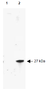
![Western blot of RFP recombinant protein detected with anti-RFP antibody. Lane 1 shows no reaction against a GFP recombinant protein present in 10 μg of HeLa cell extract. Lane 2 shows a single band detected in 10 μg of a HeLa lysate containing RFP recombinant protein. Anti-RFP detects a 27 kDa band corresponding to the epitope tag RFP. A 4-12% Bis-Tris gradient gel (Invitrogen) was used for SDS-PAGE. The protein was transferred to nitrocellulose using standard methods. After blocking the membrane was probed with the primary antibody diluted 1:2,500 for 1 h at room temperature followed by washes and reaction with a 1:5,000 dilution of IRDye(TM)800 conjugated Goat-a-Rabbit IgG [H&L]. IRDye(TM)800 fluorescence image was captured using the Odyssey® Infrared Imaging System developed by LI-COR. IRDye is a trademark of LI-COR, Inc. Other detection systems will yield similar results.](https://m1.acris-antibodies.com/catalog/product/cache/1/small_image/50x50/9df78eab33525d08d6e5fb8d27136e95/0163854.png) |
Rabbit | IgG | HRP | C, E, WB |
0.1 mg /
€515.00
|
||
| OriGene Technologies GmbH | |||||||

