RPL27A antibody
Principal name
RPL27A antibody
Alternative names for RPL27A antibody
60S ribosomal protein L27a
SwissProt ID
O14388 (Schpo), P0C2H6 (Yeast), P14115 (Mouse), P18445 (Rat), P41092 (Drome), P46776 (Human), P47830 (Xenla), P48160 (Dicdi), Q29333 (Pig), Q56K03 (Bovin)
Ncbi ID
Available reactivities
Available hosts
Available applications
Enzyme Immunoassay (E), Western blot / Immunoblot (WB), Immunocytochemistry/Immunofluorescence (ICC/IF), Paraffin Sections (P)
Background of RPL27A antibody
- Kit Component:
- - KN315032G1, Rpl27a gRNA vector 1 in pCas-Guide vector
- KN315032G2, Rpl27a gRNA vector 2 in pCas-Guide vector
- KN315032D, donor vector containing left and right homologous arms and Luciferase-Puro functional cassette
- GE100003, scramble sequence in pCas-Guide vector - CRISPR Manual:
- CRISPR/CAS9 Genome Editing Manual
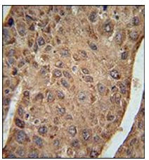
Immunohistochemistry analysis in Formalin Fixed, Paraffin Embedded Human hepatocarcinoma using RPL27A Antibody (C-term) Cat.-No. AP53714PU-N followed by peroxidase conjugation of the secondary antibody and DAB staining. This data demonstrates the use of RPL27A Antibody (C-term) for immunohistochemistry. Clinical relevance has not been evaluated.
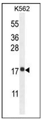
Western blot analysis in K562 cell line lysates (35 ug/lane) using RPL27A Antibody (C-term) Cat.-No. AP53714PU-N. This demonstrates the RPL27A antibody detected the RPL27A protein (arrow).
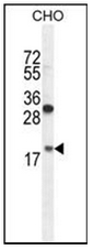
Western blot analysis in CHO cell line lysates (35 ug/lane) using RPL27A Antibody (C-term) Cat.-No. AP53714PU-N. This demonstrates the RPL27A antibody detected the RPL27A protein (arrow).
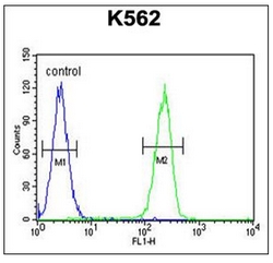
Flow Cytometric analysis of K562 cells (right histogram) compared to a negative control cell (left histogram) using RPL27A Antibody (C-term) Cat.-No. AP53714PU-N. FITC-conjugated Goat-anti-Rabbit secondary antibodies were used for the analysis.
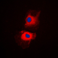
Immunofluorescent analysis of RPL27A staining in K562 cells. Formalin-fixed cells were permeabilized with 0.1% Triton X-100 in TBS for 5-10 minutes and blocked with 3% BSA-PBS for 30 minutes at room temperature. Cells were probed with the primary antibody in 3% BSA-PBS and incubated overnight at 4 C in a humidified chamber. Cells were washed with PBST and incubated with a DyLight 594-conjugated secondary antibody (red) in PBS at room temperature in the dark. DAPI was used to stain the cell nuclei (blue).
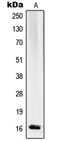
Western blot analysis of RPL27A expression in K562 (A) whole cell lysates.
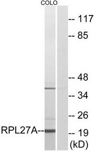
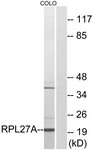
Western blot analysis of extracts from COLO cells, using RPL27A antibody.The lane on the right is treated with the synthesized peptide.
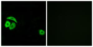
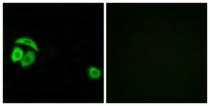
Immunofluorescence analysis of A549 cells, using RPL27A antibody.The picture on the right is treated with the synthesized peptide.
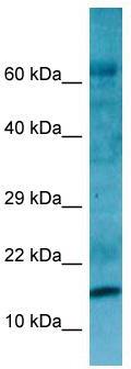
WB Suggested Anti-RPL27A Antibody; Titration: 1.0 ug/ml; Positive Control: Fetal kidney
- Quick-Filters:
- Highly Validated (1)
Primary Antibodies
| Catalog No. | Host | Iso. | Clone | Pres. | React. | Applications | |
|---|---|---|---|---|---|---|---|
| AP53714PU-N | RPL27A (C-term) antibody |
||||||
 |
Rabbit | Ig | Aff - Purified | Hst, Hu | P, WB |
0.4 ml /
€415.00
|
|
| OriGene Technologies GmbH | |||||||
 +3 additional images +3 additional images |
Highly validated antibody! |
||||||
| TA314465 | RPL27A antibody |
||||||
| Rabbit polyclonal anti-RPL27A antibody | |||||||
 |
Rabbit | IgG | Purified | Hu, Ms, Rt | E, ICC/IF, WB |
0.1 ml /
€335.00
|
|
| OriGene Technologies, Inc. | |||||||
| +1 additional image |
|||||||
Mouse Lenti ORF Particles
| Catalog No. | ||
|---|---|---|
| MR201017L2V | Lenti ORF particles, Rpl27a (GFP-tagged) - Mouse ribosomal protein L27A (Rpl27a), 200 uL, >10^7 TU/mL |
|
 |
200 µl /
€930.00
|
|
| OriGene Technologies, Inc. | ||
| MR201017L1V | Lenti ORF particles, Rpl27a (Myc-DDK-tagged) - Mouse ribosomal protein L27A (Rpl27a), 200 uL, >10^7 TU/mL |
|
|
200 µl /
€830.00
|
|
| OriGene Technologies, Inc. | ||