S100A1 antibody
Principal name
S100A1 antibody
Alternative names for S100A1 antibody
S100-A1, S100A, S100 alpha, S100 calcium-binding protein A1, S-100 protein subunit alpha, S-100 protein alpha chain
SwissProt ID
P02639 (Bovin), P23297 (Human), P35467 (Rat), P56565 (Mouse), Q5RC36 (Ponab), Q7LZT1 (Misfo)
Ncbi ID
NP_006262, NP_001092512, 9606, NP_006262.1, NP_001092512.1, NP_035439, NM_006271, NM_011309, NM_001007636
Available reactivities
Available hosts
Available applications
Enzyme Immunoassay (E), ELISA (capture) (E(capture)), Western blot / Immunoblot (WB), ELISA (detection) (E(detection)), Frozen Sections (C), Paraffin Sections (P), Immunocytochemistry/Immunofluorescence (ICC/IF), Flow Cytometry (F), Immunoprecipitation (IP)
Background of S100A1 antibody
S-100 protein derived from brain tissue is an acidic calcium-binding protein with molecular weight of about 21kDa. In human brain tissue S-100 protein is mainly presented as two isoforms - betabeta homodimer (S-100b) and alphabeta heterodimer (S-100a). Because of its predominant location in astroglial cells S-100 protein can be used as a sensitive and reliable marker for central nervous system injury. Structural damage of glial cells causes leakage of S-100 protein into the extracellular matrix and into cerebrospinal fluid, further releasing into the bloodstream. Measurements of S-100 protein in patient serum samples are useful in monitoring of traumatic brain injury, ischemic brain damage after circulatory arrests, and in diagnosis and prognosis of clinical outcome in acute stroke. Although predominant among the water-soluble brain proteins, S-100 is also found in a variety of other tissues. S-100 is an intracellular protein that weakly binds calcium. It binds zinc very tightly, however, and this appears to increase the affinity of the protein for calcium. Distinct binding sites, with different affinities, exist for both ions on each monomer. Physiological concentrations of potassium ion antagonize the binding of both divalent cations, especially affecting high-affinity calcium-binding sites.


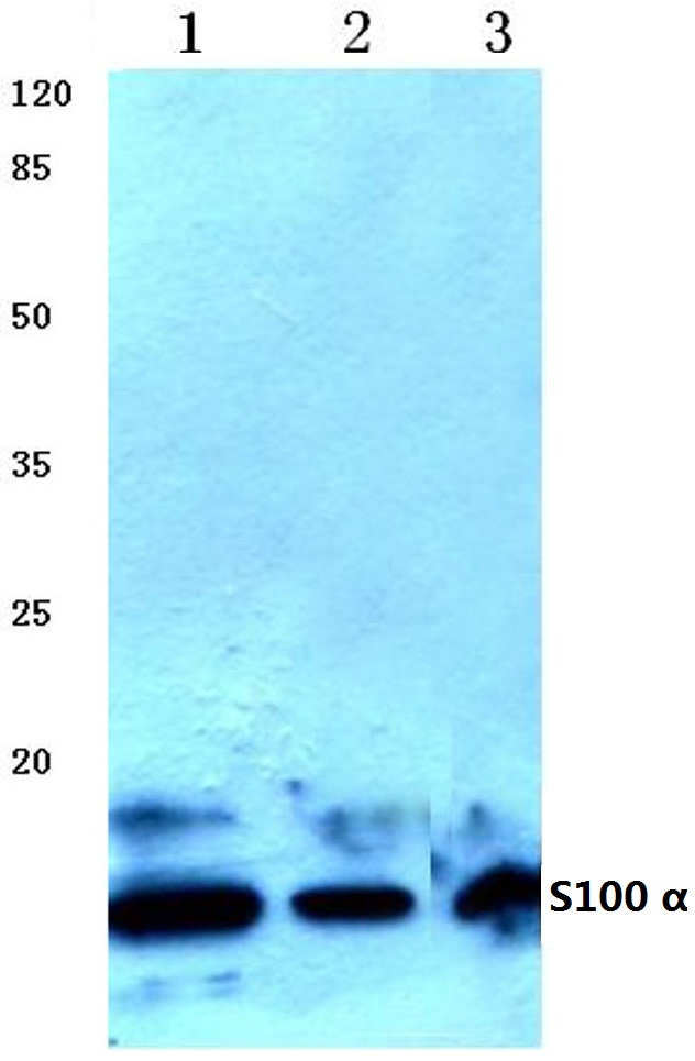
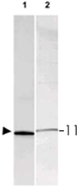
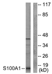
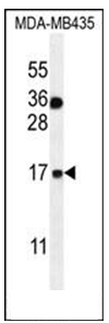
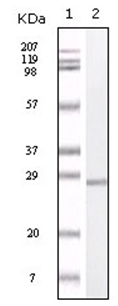
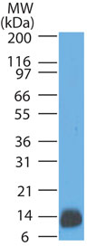
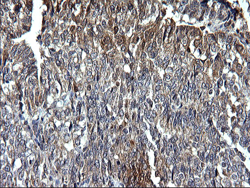
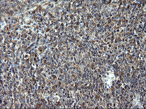
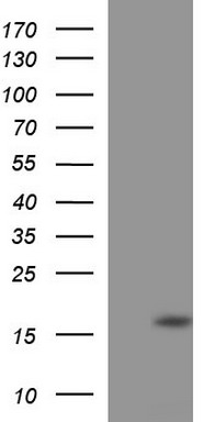
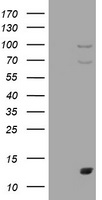
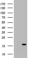
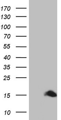
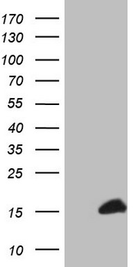
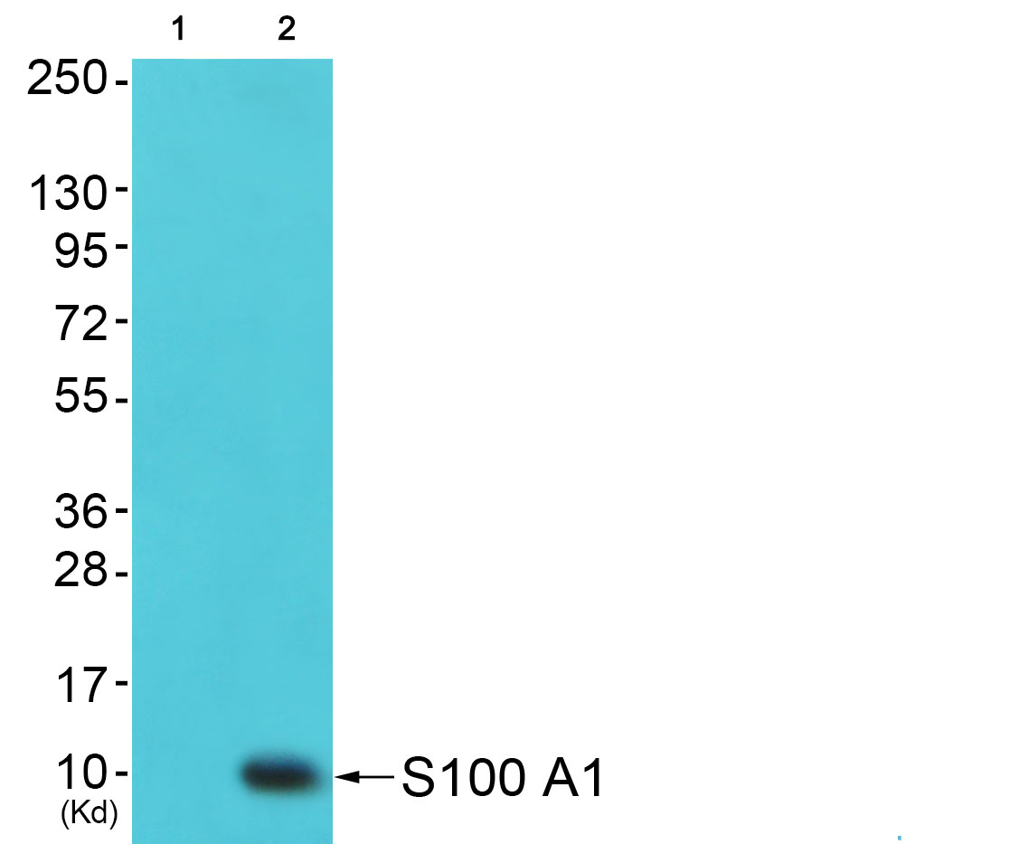
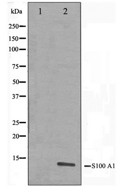
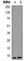
Lane1:HEK293T whole cell lysate
Lane2:Mouse brain tissue lysate
Lane3:Rat brain tissue lysate
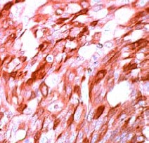
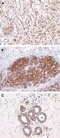
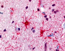
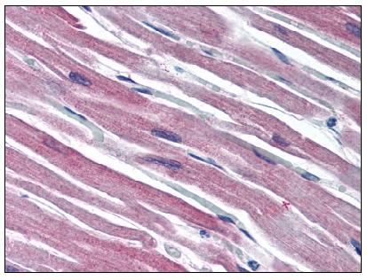
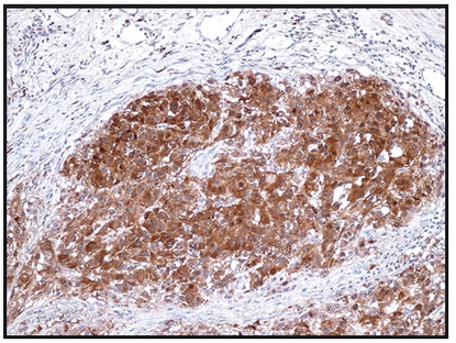
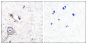
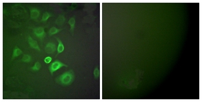









- Quick-Filters:
- Cited (2)
- Highly Validated (1)

Primary Antibodies
| Catalog No. | Host | Iso. | Clone | Pres. | React. | Applications | |
|---|---|---|---|---|---|---|---|
| AM32830PU-S | S100 antibody |
||||||
 |
Mouse | IgG2a | 4C4.9 | Purified | Bov, Hu, Ms, Rt | C, F, ICC/IF, IP, P, WB |
0.1 mg /
€430.00
|
| OriGene Technologies GmbH | |||||||
 +2 additional images +2 additional images |
Published recently! OriGene recommends this product for Immunohistochemistry. |
||||||
| AM32830PU-T | S100 antibody |
||||||
|
Mouse | IgG2a | 4C4.9 | Purified | Bov, Hu, Ms, Rt | C, F, ICC/IF, IP, P, WB |
20 µg /
€240.00
|
| OriGene Technologies GmbH | |||||||
| +2 additional images |
Published recently! OriGene recommends this product for Immunohistochemistry. |
||||||
| TA312311 | S100A1 antibody |
||||||
| Rabbit polyclonal anti-S100 A1 antibody | |||||||
 |
Rabbit | IgG | Purified | Hu, Ms, Rt | ICC/IF, WB |
0.1 ml /
€335.00
|
|
| OriGene Technologies, Inc. | |||||||
| +3 additional images |
Highly validated antibody! |
||||||
| TA319193 | S100A1 antibody |
||||||
| Rabbit polyclonal S100 Protein antibody | |||||||
![WB using Anti-S-100 antibody shows detection of a band ~11 kDa corresponding to bovine S-100 monomer (100 ng loaded, arrowhead lane 1). The antibody also detects S-100 from rat brain lysate (lane 2). The primary antibody was diluted to 1:1,000 for 2h at RT, followed by washes and reaction with a 1:10,000 dilution of IRDye™800 conjugated Gt-a-Rabbit IgG [H&L] for 45 min at room temperature.](https://m1.acris-antibodies.com/catalog/product/cache/1/small_image/50x50/9df78eab33525d08d6e5fb8d27136e95/OriGene/200-401-874-1-W.jpg) |
Rabbit | IgG | Aff - Purified | Bov | E, P, WB |
0.5 mg /
€590.00
|
|
| OriGene Technologies, Inc. | |||||||
| +2 additional images |
|||||||
| TA321757 | S100A1 antibody |
||||||
| Anti-S100A1 Rabbit Polyclonal Antibody | |||||||
 |
Rabbit | IgG | Purified | Hu, Ms, Rt | E, P |
0.1 ml /
€335.00
|
|
| OriGene Technologies, Inc. | |||||||
| TA321758 | S100A1 antibody |
||||||
| Anti-S100A1 Rabbit Polyclonal Antibody | |||||||
 |
Rabbit | IgG | Purified | Hu, Ms, Rt | E, P |
0.1 ml /
€335.00
|
|
| OriGene Technologies, Inc. | |||||||
| TA327733 | S100A1 antibody |
||||||
| Mouse Monoclonal S-100 Antibody | |||||||
 |
Mouse | IgG2a | 4C4.9 | Purified | Hu | P |
1 ml /
€445.00
|
| OriGene Technologies, Inc. | |||||||
| TA347469 | S100A1 antibody |
||||||
| Rabbit Polyclonal Anti-S100 A1 Antibody | |||||||
 |
Rabbit | IgG | Purified | Hu, Ms, Rt | WB |
0.1 mg /
€335.00
|
|
| OriGene Technologies, Inc. | |||||||
| TA349435 | S100A1 antibody |
||||||
| Rabbit Polyclonal Anti-S100A1 Antibody | |||||||
 |
Rabbit | IgG | Purified | Hu, Ms, Rt | E, P |
0.1 ml /
€335.00
|
|
| OriGene Technologies, Inc. | |||||||
| +1 additional image |
|||||||
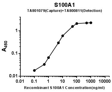
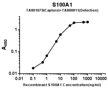
| TA800811 | S100A1 antibody |
||||||
| S100A1 (S100 alpha) mouse monoclonal antibody, clone OTI6G4 (formerly 6G4) | |||||||
 |
Mouse | IgG1 | OTI6G4 | Purified | Hu | E, WB |
0.1 ml /
€360.00
|
| OriGene Technologies, Inc. | |||||||
| +1 additional image |
|
||||||
| TA801079 | S100A1 antibody |
||||||
| S100A1 (S100 alpha) mouse monoclonal antibody, clone OTI9F3 (formerly 9F3) | |||||||
 |
Mouse | IgG1 | OTI9F3 | Purified | Hu | E, WB |
0.1 ml /
€379.00
|
| OriGene Technologies, Inc. | |||||||
| +1 additional image |
|
||||||
| TA807339 | S100A1 antibody |
||||||
| S100A1 mouse monoclonal antibody, clone OTI7B11 (formerly 7B11) | |||||||
 |
Mouse | IgG1 | OTI7B11 | Purified | Hu | P, WB |
0.1 ml /
€360.00
|
| OriGene Technologies, Inc. | |||||||
| +1 additional image |
|
||||||
| TA807374 | S100A1 antibody |
||||||
| S100A1 mouse monoclonal antibody, clone OTI2A4 (formerly 2A4) | |||||||
 |
Mouse | IgG1 | OTI2A4 | Purified | Hu | P, WB |
0.1 ml /
€360.00
|
| OriGene Technologies, Inc. | |||||||
| +1 additional image |
|
||||||
| AM06160SU-N | S100A1 antibody |
||||||
 |
Mouse | IgG1 | 2C8B8 | Ascites | Hu | P, WB |
0.1 ml /
€380.00
|
| OriGene Technologies GmbH | |||||||
| +1 additional image |
|||||||
| AM08285PU-N | S100A1 (C-term) antibody |
||||||
 |
Rabbit | Ig | Purified | Hu | P |
0.1 ml /
€425.00
|
|
| OriGene Technologies GmbH | |||||||
| AP06310PU-N | S100A1 antibody |
||||||
 |
Rabbit | Aff - Purified | Hu, Ms, Rt | E, P, WB |
0.1 mg /
€370.00
|
||
| OriGene Technologies GmbH | |||||||
| +1 additional image |
|||||||
| AP07684PU-N | S100A1 (Full Length) antibody |
||||||
 |
Rabbit | Purified | Bov, Hu | E, P, WB |
50 µg /
€425.00
|
||
| OriGene Technologies GmbH | |||||||
| AP53776PU-N | S100A1 (C-term) antibody |
||||||
 |
Rabbit | Ig | Aff - Purified | Hu | P, WB |
0.4 ml /
€415.00
|
|
| OriGene Technologies GmbH | |||||||
| +1 additional image |
|||||||
| 4S37-3B10 | S100A1 (ab) antibody |
||||||
 |
Mouse | IgG2a | 3B10 | Purified | Hu | E, E(capture), WB |
1 mg /
€420.00
|
| HyTest Ltd. | |||||||
| 4S37-6G1 | S100A1 (bb+ab) antibody |
||||||
|
Mouse | IgG1 | 6G1 | Purified | Hu | E, E(detection), WB |
1 mg /
€420.00
|
| HyTest Ltd. | |||||||