SFRP1 antibody
Principal name
SFRP1 antibody
Alternative names for SFRP1 antibody
SFRP-1, FRP-1, FRP1, FRP, SARP2, SARP-2, Secreted frizzled-related protein 1, Secreted apoptosis-related protein 2
Ncbi ID
NP_003003, NP_003003.3, 9606, AAH36503, NM_003012, NP_038862.2, NM_013834
Available reactivities
Available hosts
Available applications
Western blot / Immunoblot (WB), Paraffin Sections (P), Enzyme Immunoassay (E), Immunocytochemistry/Immunofluorescence (ICC/IF), Flow Cytometry (F)
Background of SFRP1 antibody
- Kit Component:
- - KN315654G1, Sfrp1 gRNA vector 1 in pCas-Guide vector
- KN315654G2, Sfrp1 gRNA vector 2 in pCas-Guide vector
- KN315654D, donor vector containing left and right homologous arms and Luciferase-Puro functional cassette
- GE100003, scramble sequence in pCas-Guide vector - CRISPR Manual:
- CRISPR/CAS9 Genome Editing Manual
General readings
Caldwell,G.M., et al., (2006) Br. J. Cancer 94 (6), 922-927
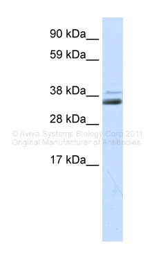
Human kidney; WB Suggested Anti-SFRP1 Antibody Titration: 0.3ug/ml. ELISA Titer: 1:62500. Positive Control: Human kidney; SFRP1 antibody - middle region (AVARP09053_T100) in Human kidney cells using Western Blot

Figure 1. Formalin-Fixed Paraffin-Embedded (FFPE) on Heart.
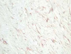
Immunohistochemistry of SFRP1 polyclonal antibody ( Cat # PAB10235 ) was used at a 1 : 800 dilution for 20 min to detect SFRP1 in human dermal hypertrophic scar tissue. Tissue was formalin-fixed followed by heat mediated antigen retrieval prior to blocking. HRP Goat-anti-Rabbit IgG ( p/n 611-1302 ) is suitable for secondary antibody detection.
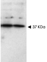
Western blot using SFRP1 polyclonal antibody ( Cat # PAB10235 ) shows detection of a band ~37 kDa band ( arrowhead ) corresponding to SFRP1 in lysates from human cultured airway epithelial cells.Lysates were run on a SDS-PAGE and transferred onto nitrocellulose followed by reaction with a 1:230 dilution of SFRP1 polyclonal antibody overnight at 4°C.Signal was detected using standard techniques.Personnel communication Becky Mercer, University of Columbia.

AP22384PU-N SFRP1 Antibody staining of Formalin-Fixed, Paraffin-Embedded Human Kidney. Steamed antigen retrieval with citrate buffer pH 6, AP-staining.

AP23107PU-N SFRP1 Antibody staining of Formalin-Fixed, Paraffin-Embedded HUman Uterus at 3.75 µg/ml after heat-induced antigen retrieval.

AP23107PU-N SFRP1 Antibody staining of Formalin-Fixed, Paraffin-Embedded HUman Kidney at 3.75 µg/ml after heat-induced antigen retrieval.
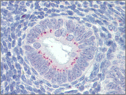
Uterus, Human: Formalin-Fixed, Paraffin-Embedded (FFPE)
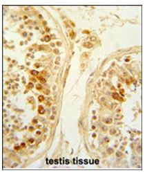
Formalin-Fixed and paraffin-Embedded Human testis tissue reacted with SFRP1 Antibody (N-term), which was peroxidase-conjugated to the secondary antibody, followed by DAB staining. This data demonstrates the use of this antibody for immunohistochemistry; clinical relevance has not been evaluated.
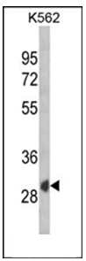
Western blot analysis of SFRP1 Antibody (N-term) Cat.-No AP53873PU-N in K562 cell line lysates (35ug/lane). SFRP1 (arrow) was detected using the purified Pab.
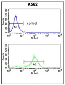
Flow Cytometry analysis of K562 cells using SFRP1 Antibody (N-term) Cat.-No AP53873PU-N (Bottom histogram) compared to a negative control cell (top histogram). FITC-conjugated Goat-anti-Rabbit secondary antibodies were used for the analysis.
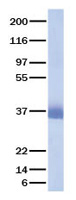
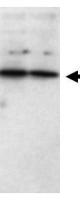
Western blot using Affinity Purified anti-SFRP1 antibody shows detection of a band ~37 kDa (arrowhead) corresponding to SFRP1 in lysates from human cultured airway epithelial cells. Lysates were run on a SDS-PAGE and transferred onto nitrocellulose followed by reaction with a 1:230 dilution of anti-SFRP1 antibody overnight at 4°C. Signal was detected using standard techniques. Personnel communication Becky Mercer, University of Columbia.

Affinity Purified anti-Human SFRP1 antibody was used at a 1:800 dilution for 20 min to detect SFRP in human dermal hypertrophic scar tissue. Tissue was formalin-fixed followed by heat mediated antigen retrieval prior to blocking. HRP Gt-a-Rabbit IgG (p/n 611-1302) is suitable for secondary antibody detection.
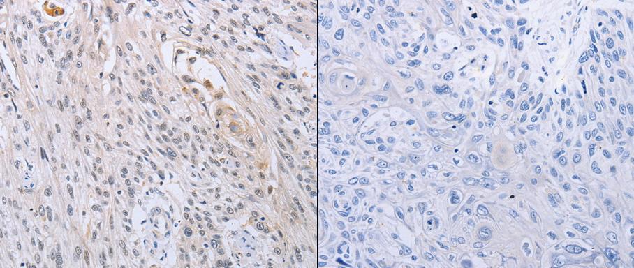
Predicted cell location: Cytoplasm. Positive control: Human esophagus cancer tissue. Recommended dilution: 1/10-50 The image on the left is immunohistochemistry of paraffin-embedded Human esophagus cancer tissue using SFRP1 antibody at dilution 1/15, on the right is treated with the synthetic peptide. (Original magnification:x200)
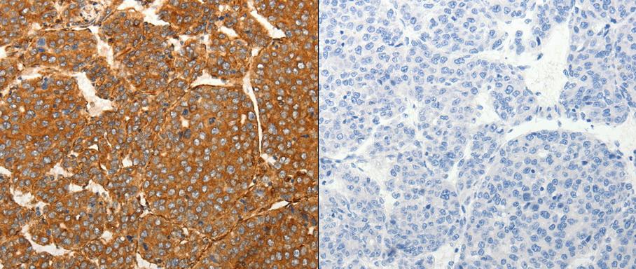
Predicted cell location: Cytoplasm. Positive control: Human liver and lung cancer tissue . Recommended dilution: 1/100-300 The image on the left is immunohistochemistry of paraffin-embedded human liver cancer tissue using SFRP1 antibody at dilution 1/80, on the right is treated with the synthetic peptide. (Original magnification:x 200)
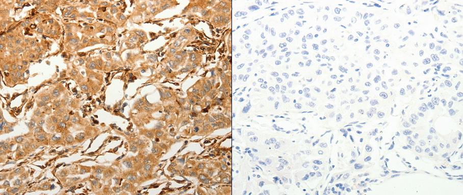
Predicted cell location: Cytoplasm. Positive control: Human liver and lung cancer tissue . Recommended dilution: 1/100-300 The image on the left is immunohistochemistry of paraffin-embedded human lung cancer tissue using SFRP1 antibody at dilution 1/80, on the right is treated with the synthetic peptide. (Original magnification:x200)

WB Suggested Anti-SFRP1 Antibody Titration: 0.3 ug/ml; ELISA Titer: 1:62500; Positive Control: Human kidney
Primary Antibodies
| Catalog No. | Host | Iso. | Clone | Pres. | React. | Applications | |
|---|---|---|---|---|---|---|---|
| AP53873PU-N | SFRP1 (N-term) antibody |
||||||
 |
Rabbit | Ig | Aff - Purified | Hu | F, P, WB |
0.4 ml /
€415.00
|
|
| OriGene Technologies GmbH | |||||||
 +2 additional images +2 additional images |
|||||||