SMAD4 antibody
Principal name
SMAD4 antibody
Alternative names for SMAD4 antibody
SMAD family member 4, SMAD-4, SMAD 4, MADH4, MAD homolog 4, DPC4, Mothers against decapentaplegic homolog 4, Mothers against DPP homolog 4, Deletion target in pancreatic carcinoma 4
Ncbi ID
NP_005350.1, 9606, NP_005350, 10090, ABM82766.1, NM_008540, NM_019275, NM_005359
Available reactivities
Available hosts
Available applications
Western blot / Immunoblot (WB), Enzyme Immunoassay (E), Immunocytochemistry/Immunofluorescence (ICC/IF), Paraffin Sections (P), Dot blot (Dot), Immunoprecipitation (IP)
Background of SMAD4 antibody
SMAD4 (also known as Mothers against decapentaplegic homolog 4, Mothers against DPP homolog 4, deletion target in pancreatic carcinoma 4 and hSMAD4) is a common mediator of signal transduction by TGF-beta (transforming growth factor), but is also involved in cancer development and metastases as a tumor suppressor. SMAD4 promotes binding of the SMAD2/SMAD4/FAST-1 complex to DNA and provides an activation function required for SMAD1 or SMAD2 to stimulate transcription. SMAD4 may form trimers with receptor-regulated SMAD (R-SMAD) and interacts with ATF2, COPS5, DACH1, MSG1, SKI and TRIM33. In the absence of ligand SMAD4 is found in the cytoplasm, but when complexed with R-SMAD, translocates to the nucleus. Defects in SMAD4 are a cause of pancreatic carcinoma and juvenile polyposis syndrome (JPS), a syndrome in which patients are at risk for developing gastrointestinal cancers.
General readings
1. Kim, B.-G.; Li, C.; Qiao, W.; Mamura, M.; Kasprzak, B.; Anver, M.; Wolfraim, L.; Hong, S.; Mushinski, E.; Potter, M.; Kim, S.-J.; Fu, X.-Y.; Deng, C.; Letterio, J. J. : Smad4 signalling in T cells is required for suppression of gastrointestinal cancer. Nature 441: 1015-1019, 2006. Note: Erratum: Nature 444: 780 only, 2006.
2. Houlston, R.; Bevan, S.; Williams, A.; Young, J.; Dunlop, M.; Rozen, P.; Eng, C.; Markie, D.; Woodford-Richens, K.; Rodriguez-Bigas, M. A.; Leggett, B.; Neale, K.; Phillips, R.; Sheridan, E.; Hodgson, S.; Iwama, T.; Eccles, D.; Bodmer, W.; Tomlinson, I. : Mutations in DPC4 (SMAD4) cause juvenile polyposis syndrome, but only account for a minority of cases. Hum. Molec. Genet. 7: 1907-1912, 1998.
3 Howe, J. R.; Roth, S.; Ringold, J. C.; Summers, R. W.; Jarvinen, H. J.; Sistonen, P.; Tomlinson, I. P. M.; Houlston, R. S.; Bevan, S.; Mitros, F. A.; Stone, E. M.; Aaltonen, L. A. : Mutations in the SMAD4/DPC4 gene in juvenile polyposis. Science 280: 1086-1088, 1998.
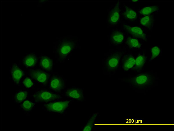
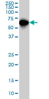
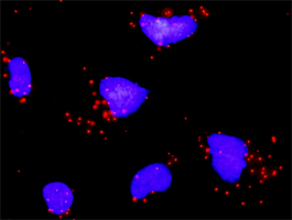
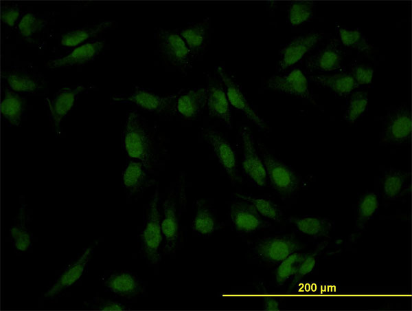
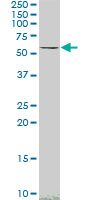

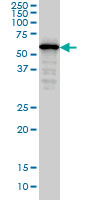
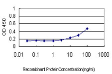
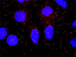

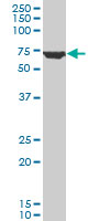


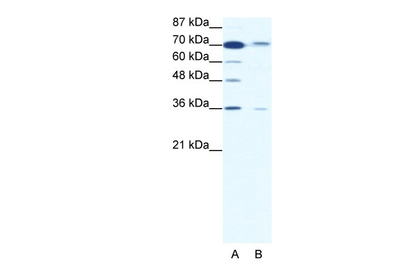
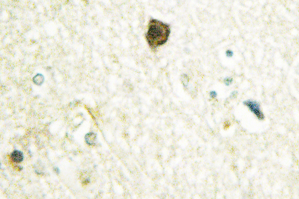
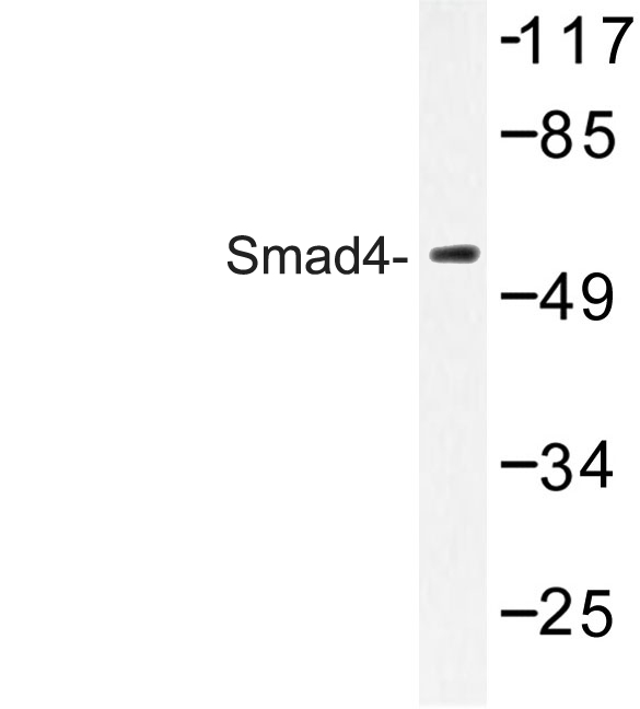
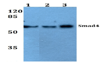
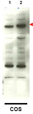
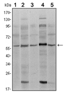
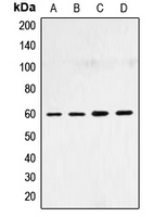
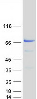
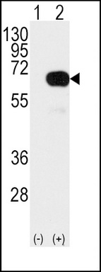
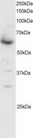
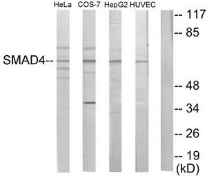
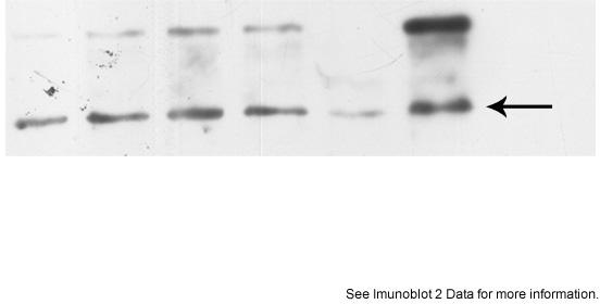
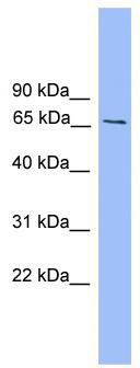
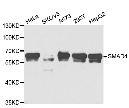
Lane 1: A549 whole cell lysate.
Lane 2: Raw264.7 whole cell lysate.
Lane 3: PC12 whole cell lysate.


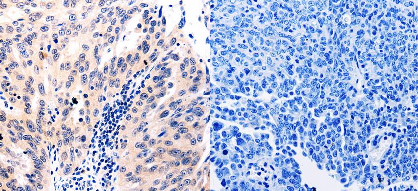
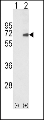

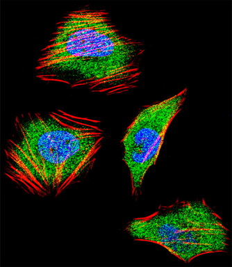
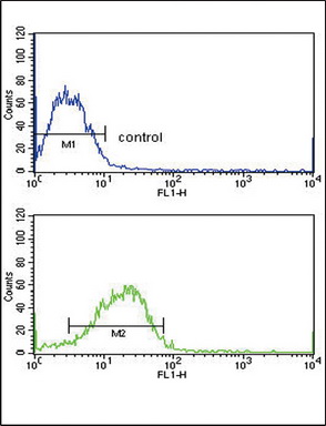

- Quick-Filters:
- Highly Validated (1)
Primary Antibodies
| Catalog No. | Host | Iso. | Clone | Pres. | React. | Applications | |
|---|---|---|---|---|---|---|---|
| AM06501SU-N | SMAD4 antibody |
||||||
 |
Mouse | IgG1 | 4G1C6 | Ascites | Hu | E, ICC/IF, P, WB |
0.1 ml /
€380.00
|
| OriGene Technologies GmbH | |||||||
 +3 additional images +3 additional images |
Highly validated antibody! |
||||||
| TA302724 | SMAD4 (C-term) antibody |
||||||
| Goat Polyclonal Antibody against SMAD4 | |||||||
 |
Goat | Aff - Purified | Bov, Can, Hu, Ms, Por, Rt | E, WB |
0.1 mg /
€360.00
|
||
| OriGene Technologies, Inc. | |||||||
| TA321708 | SMAD4 antibody |
||||||
| Anti-SMAD4 Rabbit Polyclonal Antibody | |||||||
 |
Rabbit | IgG | Purified | Hu, Ms, Rt | E, P |
0.1 ml /
€335.00
|
|
| OriGene Technologies, Inc. | |||||||
| AP09377PU-N | SMAD4 antibody |
||||||
 |
Rabbit | IgG | Aff - Purified | Hu, Ms, Xen | E, WB |
0.1 mg /
€515.00
|
|
| OriGene Technologies GmbH | |||||||