SOCS1 antibody
Principal name
SOCS1 antibody
Alternative names for SOCS1 antibody
Suppressor of cytokine signaling 1, SOCS-1, SSI1, SSI-1, TIP3, TIP-3, JAB, JAK-binding protein, STAT-induced STAT inhibitor 1, Tec-interacting protein 3
Ncbi ID
9606, NP_003736, NP_003736.1, 10116, NM_003745, AAI53089.1, CAB92528, NM_009896, NM_145879
Available reactivities
Available hosts
Available applications
Western blot / Immunoblot (WB), Enzyme Immunoassay (E), Paraffin Sections (P), Frozen Sections (C), Immunoprecipitation (IP), Immunocytochemistry/Immunofluorescence (ICC/IF)
Background of SOCS1 antibody
Suppressor of cytokine signaling 1 (SOCS1) is a member of the SOCS family of proteins, consisting of eight members (SOCS1 to SOCS7 and CIS) all sharing a central SH2 domain and a c-terminal SOCS box (1). This family is involved in inducible feedback inhibition of janus kinases (JAKs) and signal transducers and activators of transcription (STATs) signaling pathway, which plays multiple roles in cell growth, differentiation, and apoptosis (2). SOCS1 is located on chromosome 16 (3). It plays a key role in the negative regulation of IFN-gamma signaling and in T-cell differentiation. SOCS1 inhibits macrophage differentiation of M1 cells in response to IL-6, protects protein tyrosine phosphatases by thioredoxin upregulation and attenuates JAKs to suppress ROS-mediated apoptosis (2, 4). It translocates into the nucleus due to the presence of a functional nuclear localization signal (NLS). It also regulates IFN-gamma mediated sensory neuronal survival.
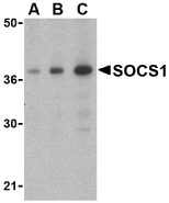
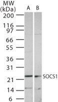
SOCS1 is implicated in various cancers and diseases. Mutations of SOCS1 are found in classic Hodgkin lymphoma and primary mediastinal B-cell lymphoma (5). It is induced in response to TNF-alpha or H2O2 and by a subset of cytokines including those belonging to interferon, interleukin and colony stimulating factor colonies. It attenuates their signaling by its ability to bind and inhibit all four of the JAKs. It is found to colocalize and biochemically copurify with the microtubule organizing complex (MTOC) and its associated 20S proteasome (6). It is expressed in all tissues with high expression in spleen, small intestine and peripheral blood lymphocytes. It is approximately a 25kDa protein (211 amino acids).
General readings
1. Larsen L, Ropke C. Suppressors of Cytokine Signaling: SOCS. APMIS. December 2002; 110(12):833-844.
2. Oh J, Hur MW, Lee CE. SOCS1 protects protein tyrosine phosphatases by thioredoxin upregulation and attenuates JAK to suppress ROS mediated apoptosis. Oncogene. 3 September 2009; 28(35):3145-3156.
3. Yandava CN, Pillari A, Drazen JM. Radiation hybrid and cytogenetic mapping of SOCS1 and SOCS2 to chromosomes 16p13 and 12q, respectively. Genomics. 1 October 1999; 61(1):108-111.
4. Starr R, Wilson TA, Viney EM, Murray LJ, Rayner JR, Jenkins BJ, Gonda TJ, Alexander WS, Metcalf D, Nicola NA, Hilton DJ. A family of cytokine-inducible inhibitors of signaling. Nature. 26 June 1997; 6636:917-921.
5. Weniger MA, Melzner I, Menz CK, Wegener S, Bucur AJ, Dorsch K, Mattfeldt T, Barth TF, Muller P. Mutations of the tumor suppressor gene SOCS-1 in classical Hodgkin lymphoma are frequent and associated with delayed degradation of JAK2 protein. Oncogene. 2006; 25(18):2679-2684.
6. Vuong BQ, Arenzana TL, Showalter BM, Losman J, Chen XP, Mostecki J, Banks AS, Limnander A, Fernandez N, Rothman PB. SOCS-1 localizes to the microtubule organizing complexassociated
20S proteasome. Mol Cell Biol. 2004; 24(20):9092-9101.

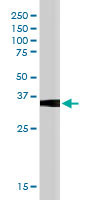
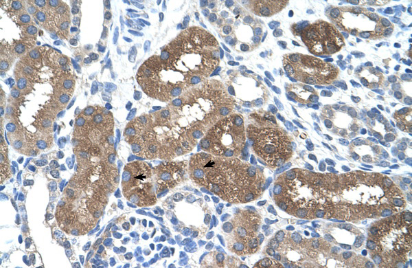

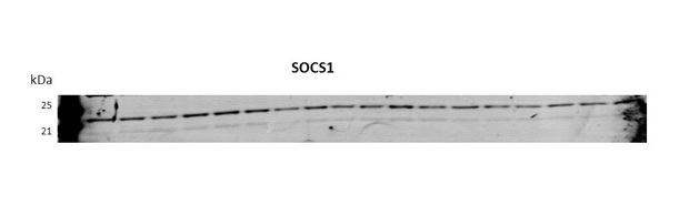

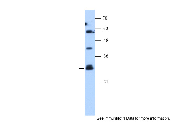
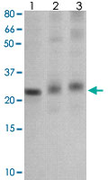
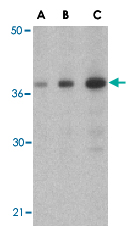
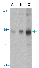


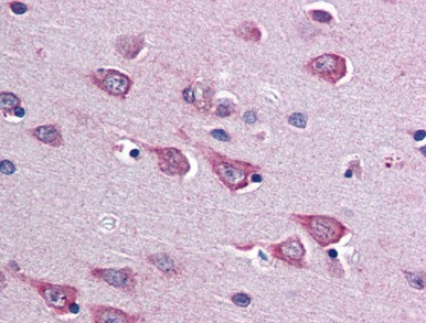

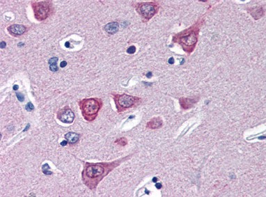
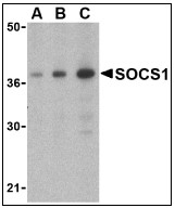
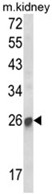

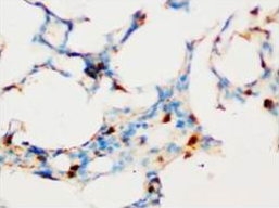
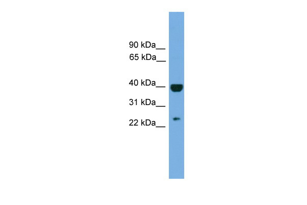
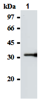
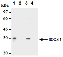
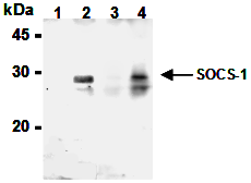
Immunoprecipitation of SOCS-1 from mouse splenocytes (1) and mouse thymocytes (3) with AM26561AF-N (1,3) or mouse IgG1 (2,4). After immunoprecipitated with the antibody, immunocomplex was resolved on SDS-PAGE and immunoblotted with biotinylated anti-SOCS-1 monoclonal antibody.



Proteins & Growth Factors
| Catalog No. | Species | Pres. | Purity | Source | |
|---|---|---|---|---|---|
| TP320847 | SOCS1 |
||||
| Recombinant protein of human suppressor of cytokine signaling 1 (SOCS1) | |||||
 |
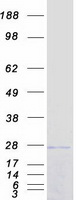
Human | > 80 % Preparation: Recombint protein was captured through anti-DDK affinity column followed by conventiol chromatography steps. Purity Detail: > 80% as determined by SDS-PAGE and Coomassie blue staining. |
HEK293 cells |
20 µg /
€748.00
|
|
| OriGene Technologies, Inc. | |||||