Splicing factor 1 (SF1) antibody
Principal name
Splicing factor 1 (SF1) antibody
Alternative names for Splicing factor 1 (SF1) antibody
ZFM1, ZNF162, BBP, Zinc finger protein 162, Transcription factor ZFM1, Zinc finger gene in MEN1 locus, Mammalian branch point-binding protein
Gene ID
7536 (SF1)
Ncbi ID
9606, NP_004621, NP_001171501.1, NP_001171501, NM_004630, AAH08080, NP_973727, NM_201998, NM_201995, NM_201997, NM_001178031, NM_001178030
Available reactivities
Available hosts
Available applications
Enzyme Immunoassay (E), Immunocytochemistry/Immunofluorescence (ICC/IF), Western blot / Immunoblot (WB), Paraffin Sections (P)
Background of Splicing factor 1 (SF1) antibody
- Kit Component:
- - KN205846G1, SF1 gRNA vector 1 in pCas-Guide vector
- KN205846G2, SF1 gRNA vector 2 in pCas-Guide vector
- KN205846D, donor vector containing left and right homologous arms and Luciferase-Puro functional cassette
- GE100003, scramble sequence in pCas-Guide vector - CRISPR Manual:
- CRISPR/CAS9 Genome Editing Manual
General readings
Huang,Y.H., (2006) Endocrinology 147 (5), 2481-2489

Immunoperoxidase of monoclonal antibody to SF1 on formalin-fixed paraffin-embedded human colon. [antibody concentration 3 ug/ml]
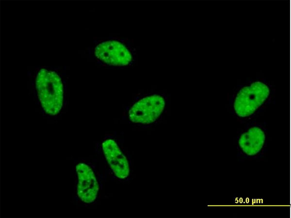
Immunofluorescence of monoclonal antibody to SF1 on HeLa cell . [antibody concentration 10 ug/ml]
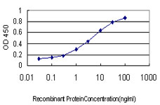
Detection limit for recombinant GST tagged SF1 is approximately 0.03ng/ml as a capture antibody.

Human Heart; Rabbit Anti-SF1 antibody. Catalog Number: ARP41214 . Paraffin Embedded Tissue: Human Heart cell Cellular Data: cardiac cell of renal tubule. Antibody Concentration: 4.0-8.0 ug/ml. Magnification: 400X; SF1 antibody - N-terminal region (ARP41214_T100) in Human Heart using Immunohistochemistry

Human Kidney; Rabbit Anti-SF1 Antibody. Catalog Number: ARP41214. Paraffin Embedded Tissue: Human Kidney. Cellular Data: Epithelial cells of renal tubule. Antibody Concentration: 4.0-8.0 ug/ml. Magnification: 400X; SF1 antibody - N-terminal region (ARP41214_T100) in Human Kidney cells using Immunohistochemistry
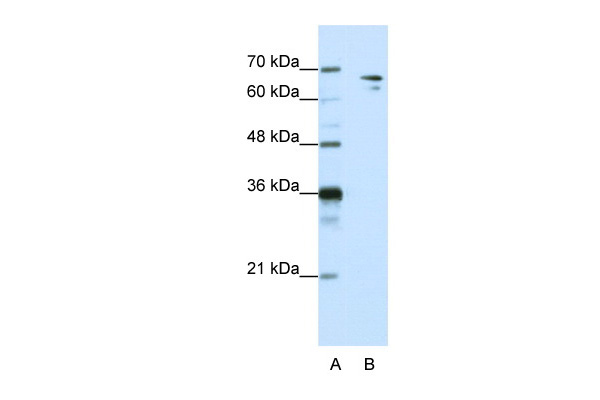
Human HepG2; WB Suggested Anti-SF1 Antibody Titration: 1.25ug/ml. ELISA Titer: 1:312500. Positive Control: HepG2 cell lysate. SF1 is supported by BioGPS gene expression data to be expressed in HepG2.; SF1 antibody - N-terminal region (ARP41214_T100) in Human HepG2 cells using Western Blot

Human HepG2; WB Suggested Anti-SF1 Antibody Titration: 0.2-1 ug/ml. ELISA Titer: 1:62500. Positive Control: HepG2 cell lysate. SF1 is supported by BioGPS gene expression data to be expressed in HepG2.; SF1 antibody - C-terminal region (ARP41212_P050) in Human HepG2 cells using Western Blot
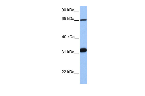
Human Jurkat; WB Suggested Anti-SF1 Antibody Titration: 0.2-1 ug/ml. Positive Control: Jurkat cell lysate. SF1 is supported by BioGPS gene expression data to be expressed in Jurkat.; SF1 antibody - C-terminal region (ARP41213_P050) in Human Jurkat cells using Western Blot
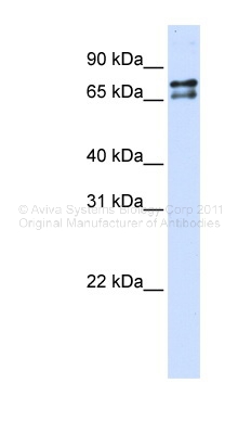
Transfected 293T; WB Suggested Anti-SF1 Antibody Titration: 0.2-1 ug/ml. ELISA Titer: 1:312500. Positive Control: Transfected 293T; SF1 antibody - N-terminal region (ARP38504_P050) in Transfected 293T cells using Western Blot
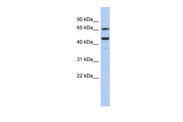
Human COLO205; WB Suggested Anti-SF1 Antibody Titration: 0.2-1 ug/ml. ELISA Titer: 1:12500. Positive Control: COLO205 cell lysate. SF1 is supported by BioGPS gene expression data to be expressed in COLO205.; SF1 antibody - middle region (ARP33493_P050) in Human COLO205 cells using Western Blot

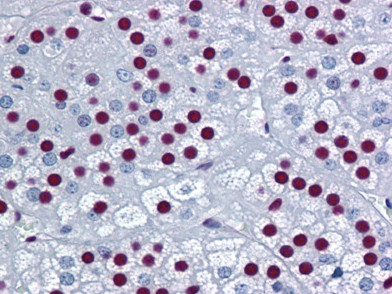
Human Adrenal: Formalin-Fixed, Paraffin-Embedded (FFPE)
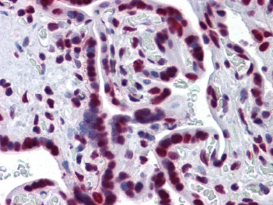
Human Placenta: Formalin-Fixed, Paraffin-Embedded (FFPE)
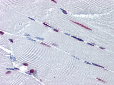
Human Skeletal Muscle: Formalin-Fixed, Paraffin-Embedded (FFPE)
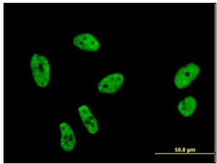
Immunofluorescence of monoclonal antibody to SF1 on HeLa cell (antibody concentration 10 µg/ml).
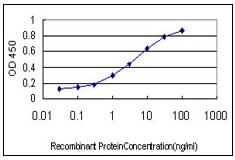
Detection limit for recombinant GST tagged SF1 is approximately 0.03 ng/ml as a capture antibody.
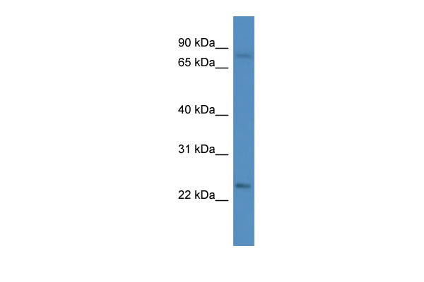
Human Jurkat; WB Suggested Anti-SF1 Antibody. . Titration: 1.0 ug/ml. . Positive Control: Jurkat Whole Cell. SF1 is supported by BioGPS gene expression data to be expressed in Jurkat.; SF1 antibody - N-terminal region (ARP33492_P050) in Human Jurkat cells using Western Blot
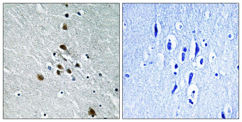
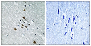
Immunohistochemical analysis of paraffin-embedded human brain tissue using SF1 (Phospho-Ser82) antibody AP55704PU-N (left)or the same antibody preincubated with blocking peptide (right).
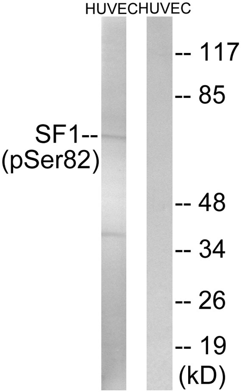
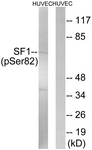
Western blot analysis of extracts from HUVEC cells treated with anisomycin using SF1 (Phospho-Ser82) Antibody AP55704PU-N.The lane on the right is treated with the antigen-specific peptide.

Western blot analysis of SF1 (pS82) expression in SHSY5Y (A); Jurkat (B) whole cell lysates.
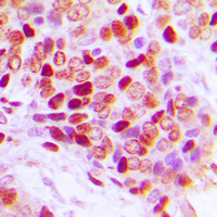
Immunohistochemical analysis of SF1 (pS82) staining in human breast cancer formalin fixed paraffin embedded tissue section. The section was pre-treated using heat mediated antigen retrieval with sodium citrate buffer (pH 6.0). The section was then incubated with the antibody at room temperature and detected using an HRP conjugad compact polymer system. DAB was used as the chromogen. The section was then counterstained with haematoxylin and mounted with DPX.
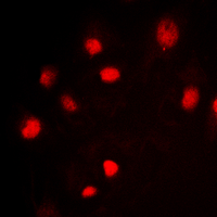
Immunofluorescent analysis of SF1 (pS82) staining in Jurkat cells. Formalin-fixed cells were permeabilized with 0.1% Triton X-100 in TBS for 5-10 minutes and blocked with 3% BSA-PBS for 30 minutes at room temperature. Cells were probed with the primary antibody in 3% BSA-PBS and incubated overnight at 4 deg C in a hidified chamber. Cells were washed with PBST and incubated with a DyLight 594-conjugated secondary antibody (red) in PBS at room temperature in the dark. DAPI was used to stain the cell nuclei (blue).
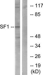
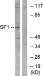
Western blot analysis of extracts from COLO205 cells, using SF1 (Ab-82) antibody.The lane on the right is treated with the synthesized peptide.

Immunohistochemistry analysis of paraffin-embedded human brain tissue using SF1 (Ab-82) antibody.The picture on the right is treated with the synthesized peptide.

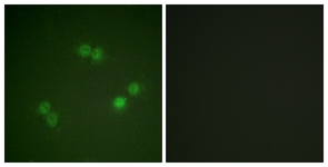
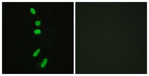
Immunofluorescence analysis of HeLa cells, using SF1 (Ab-82) antibody.The picture on the right is treated with the synthesized peptide.
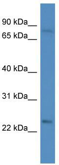
WB Suggested Anti-SF1 Antibody; Titration: 1.0 ug/ml; Positive Control: Jurkat Whole CellSF1 is supported by BioGPS gene expression data to be expressed in Jurkat
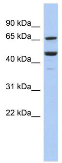
WB Suggested Anti-SF1 Antibody Titration: 0.2-1 ug/ml; ELISA Titer: 1:12500; Positive Control: COLO205 cell lysateSF1 is supported by BioGPS gene expression data to be expressed in COLO205
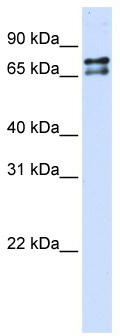
WB Suggested Anti-SF1 Antibody Titration: 0.2-1 ug/ml; ELISA Titer: 1: 312500; Positive Control: Transfected 293T
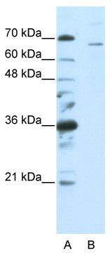
WB Suggested Anti-SF1 Antibody Titration: 0.2-1 ug/ml; ELISA Titer: 1: 62500; Positive Control: HepG2 cell lysateSF1 is supported by BioGPS gene expression data to be expressed in HepG2
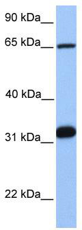
WB Suggested Anti-SF1 Antibody Titration: 0.2-1 ug/ml; Positive Control: Jurkat cell lysateSF1 is supported by BioGPS gene expression data to be expressed in Jurkat
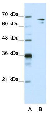
WB Suggested Anti-SF1 Antibody Titration: 1.25 ug/ml; ELISA Titer: 1: 312500; Positive Control: HepG2 cell lysateSF1 is supported by BioGPS gene expression data to be expressed in HepG2

Rabbit Anti-SF1 Antibody; Paraffin Embedded Tissue: Human Kidney; Cellular Data: Epithelial cells of renal tubule; Antibody Concentration: 4.0-8.0 ug/ml; Magnification: 400X
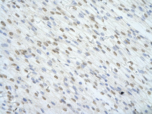
Rabbit Anti-SF1 antibody; Paraffin Embedded Tissue: Human Heart cell Cellular Data: cardiac cell of renal tubule; Antibody Concentration: 4.0-8.0 ug/ml; Magnification: 400X
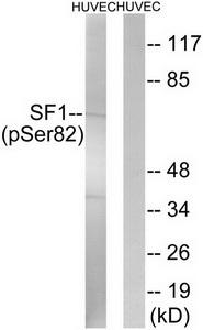
Western blot analysis of extracts from HUVEC cells, treated with anisomycin (25ug/ml, 30mins), using SF1 (Phospho-Ser82) antibody.The lane on the right is treated with the synthesized peptide.
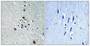
Immunohistochemistry analysis of paraffin-embedded human brain tissue using SF1 (Phospho-Ser82) antibodyThe picture on the right is treated with the synthesized peptide.
Primary Antibodies
| Catalog No. | Host | Iso. | Clone | Pres. | React. | Applications | |
|---|---|---|---|---|---|---|---|
| TA314682 | Splicing factor 1 (SF1) antibody |
||||||
| Rabbit polyclonal SF1 (Ab-82) antibody | |||||||
 |
Rabbit | IgG | Purified | Hu, Ms | E, ICC/IF, P, WB |
0.1 ml /
€335.00
|
|
| OriGene Technologies, Inc. | |||||||
 +2 additional images +2 additional images |
|||||||
| TA314683 | Splicing factor 1 (SF1) antibody |
||||||
| Rabbit polyclonal SF1 (Ser82) antibody(Phospho-specific) | |||||||
 |
Rabbit | IgG | Purified | Hu, Ms | E, ICC/IF, P, WB |
0.1 ml /
€335.00
|
|
| OriGene Technologies, Inc. | |||||||
| +1 additional image |
|||||||
| AP55704PU-N | Splicing factor 1 (SF1) pSer82 antibody |
||||||
 |
Rabbit | Aff - Purified | Hu, Ms | P, WB |
0.1 mg /
€300.00
|
||
| OriGene Technologies GmbH | |||||||
| +1 additional image |
|||||||
| AP55704PU-S | Splicing factor 1 (SF1) pSer82 antibody |
||||||
|
Rabbit | Aff - Purified | Hu, Ms | P, WB |
50 µg /
€230.00
|
||
| OriGene Technologies GmbH | |||||||
| +1 additional image |
|||||||
Proteins & Growth Factors
| Catalog No. | Species | Pres. | Purity | Source | |
|---|---|---|---|---|---|
| TP305846 | Splicing factor 1 (SF1) (transcript variant 3) |
||||
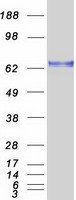
| Recombinant protein of human splicing factor 1 (SF1), transcript variant 3 | |||||
 |
Human | > 80 % Preparation: Recombint protein was captured through anti-DDK affinity column followed by conventiol chromatography steps. Purity Detail: > 80% as determined by SDS-PAGE and Coomassie blue staining. |
HEK293 cells |
20 µg /
€748.00
|
|
| OriGene Technologies, Inc. | |||||