swi6 antibody
Principal name
swi6 antibody
Alternative names for swi6 antibody
Chromatin-associated protein swi6
SwissProt ID
P40381 (Schpo)
Gene ID
2541633 (swi6)
Ncbi ID
Available reactivities
Available hosts
Available applications
Enzyme Immunoassay (E), Western blot / Immunoblot (WB), Immunoprecipitation (IP)
Background of swi6 antibody
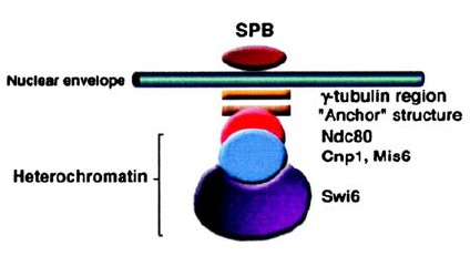
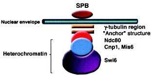
The chromatin structure of Fission yeast centromeres is regulated epigenetically. In the central core domain, the histone H3 variant Cnp1(CENP-A) plays a key role. In the flanking heterochromatin domain, histones are kept underacetylated by the histone deacetylases (HDACs) Clr3, Clr6 and Sir2, and methylated by Clr4 methyltransferase (HMTase) to create a specific binding site for the Swi6 protein. Swi6 then directly mediates cohesin binding to the centromeric heterochromatin. Recognizes and binds histone H3 tails methylated at Lys-9, leading to epigenetic repression. Involved in the repression of the silent mating-type loci MAT2 and MAT3. May compact MAT2/3 into a heterochromatin-like conformation which represses the transcription of these silent cassettes. Swi6 interacts with histone H3 methylated at Lys-9 and with the cohesin subunit psc3. Swi6 associates with Swi4 to activate HO and many other late G1-specific transcripts in budding yeast. In contrast, histone H3 methylated at Lys9 and its interacting protein Swi6/HP1, which define heterochromatin, coated extended domains associated with a variety of repeat elements and small islands corresponding to meiotic genes. Contains two chromo domains.
Primary Antibodies
| Catalog No. | Host | Iso. | Clone | Pres. | React. | Applications | |
|---|---|---|---|---|---|---|---|
| AP09279PU-N | swi6 (314-328) antibody |
||||||
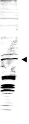
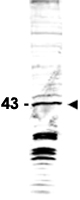
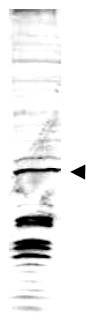
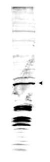
![Western blot analysis is shown using Affinity Purified anti-Swi6 antibody to detect endogenous protein present in S.pombe lysate (arrowhead). Comparison to a molecular weight marker (not shown) indicates a band of ~43 kDa corresponding to S.pombe Swi6 protein. ~35µg of lysate was loaded per lane onto a 4-20% gradient gel for SDS-PAGE followed by transfer to 0.45 µm nitrocellulose. The blot was incubated with a 1:1,700 dilution of the antibody at room temperature for 2 h followed by detection using IRDye(TM)800 labeled Goat-a-Rabbit IgG [H&L] diluted 1:5,000 for 45 min. IRDye(TM)800 fluorescence image was captured using the Odyssey(R) Infrared Imaging System developed by LI-COR. IRDye is a trademark of LI-COR, Inc. Other detection systems will yield similar results.](https://m1.acris-antibodies.com/catalog/product/cache/1/small_image/50x50/9df78eab33525d08d6e5fb8d27136e95/0164178.jpeg) |
Rabbit | IgG | Aff - Purified | Ye | E, WB |
0.1 mg /
€515.00
|
|
| OriGene Technologies GmbH | |||||||
 +1 additional image +1 additional image |
|||||||