TBC1D21 antibody
Principal name
TBC1D21 antibody
Alternative names for TBC1D21 antibody
TBC1 domain family member 21
SwissProt ID
Q8IYX1 (Human)
Gene ID
161514 (TBC1D21)
Ncbi ID
Available reactivities
Available hosts
Available applications
ELISA (detection) (E(detection)), Western blot / Immunoblot (WB), Flow Cytometry (F), Paraffin Sections (P)
Background of TBC1D21 antibody
- Kit Component:
- - KN206651G1, TBC1D21 gRNA vector 1 in pCas-Guide vector
- KN206651G2, TBC1D21 gRNA vector 2 in pCas-Guide vector
- KN206651D, donor vector containing left and right homologous arms and Luciferase-Puro functional cassette
- GE100003, scramble sequence in pCas-Guide vector - CRISPR Manual:
- CRISPR/CAS9 Genome Editing Manual
General readings
Ishibashi, K., et al. Genes Cells 14(1):41-52(2009)
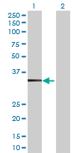
Western Blot analysis of TBC1D21 expression in transfected 293T cell line (H00161514-T01) by TBC1D21 MaxPab polyclonal antibody.Lane 1: TBC1D21 transfected lysate(36.96 KDa).Lane 2: Non-transfected lysate.
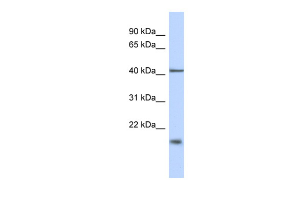
Human Brain; WB Suggested Anti-TBC1D21 Antibody Titration: 0.2-1 ug/ml. ELISA Titer: 1:1562500. Positive Control: Human brain; TBC1D21 antibody - N-terminal region (ARP53171_P050) in Human Brain cells using Western Blot
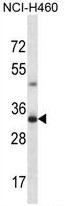
TBC1D21 Antibody (N-term) (Cat. #AP54166PU-N) western blot analysis in NCI-H460 cell line lysates (35µg/lane).This demonstrates the TBC1D21 antibody detected the TBC1D21 protein (arrow).

Immunohistochemical staining of paraffin-embedded Carcinoma of Human thyroid tissue using anti-TBC1D21 mouse monoclonal antibody. (Heat-induced epitope retrieval by 10mM citric buffer, pH6.0, 100C for 10min, TA503671)
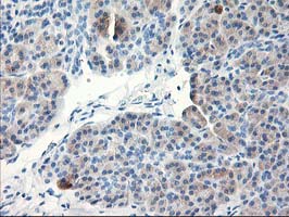
Immunohistochemical staining of paraffin-embedded Human pancreas tissue within the normal limits using anti-TBC1D21 mouse monoclonal antibody. (Heat-induced epitope retrieval by 10mM citric buffer, pH6.0, 100C for 10min, TA503671)
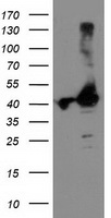
HEK293T cells were transfected with the pCMV6-ENTRY control (Left lane) or pCMV6-ENTRY TBC1D21 (RC206651, Right lane) cDNA for 48 hrs and lysed. Equivalent amounts of cell lysates (5 ug per lane) were separated by SDS-PAGE and immunoblotted with anti-TBC1D21.
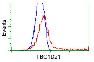
HEK293T cells transfected with either RC206651 overexpress plasmid(Red) or empty vector control plasmid(Blue) were immunostained by anti-TBC1D21 antibody(TA503671), and then analyzed by flow cytometry.
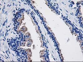
Immunohistochemical staining of paraffin-embedded Human prostate tissue within the normal limits using anti-TBC1D21 mouse monoclonal antibody. (Heat-induced epitope retrieval by 10mM citric buffer, pH6.0, 100C for 10min, TA503710)
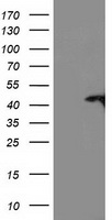
HEK293T cells were transfected with the pCMV6-ENTRY control (Left lane) or pCMV6-ENTRY TBC1D21 (RC206651, Right lane) cDNA for 48 hrs and lysed. Equivalent amounts of cell lysates (5 ug per lane) were separated by SDS-PAGE and immunoblotted with anti-TBC1D21.
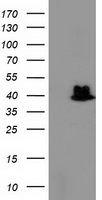
HEK293T cells were transfected with the pCMV6-ENTRY control (Left lane) or pCMV6-ENTRY TBC1D21 (RC206651, Right lane) cDNA for 48 hrs and lysed. Equivalent amounts of cell lysates (5 ug per lane) were separated by SDS-PAGE and immunoblotted with anti-TBC1D21.
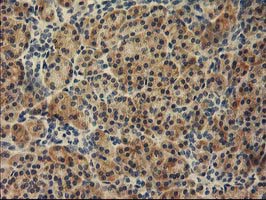
Immunohistochemical staining of paraffin-embedded Human pancreas tissue within the normal limits using anti-TBC1D21 mouse monoclonal antibody. (Heat-induced epitope retrieval by 10mM citric buffer, pH6.0, 100C for 10min, TA503718)
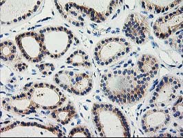
Immunohistochemical staining of paraffin-embedded Carcinoma of Human thyroid tissue using anti-TBC1D21 mouse monoclonal antibody. (Heat-induced epitope retrieval by 10mM citric buffer, pH6.0, 100C for 10min, TA503718)
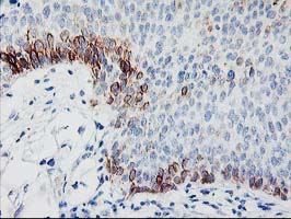
Immunohistochemical staining of paraffin-embedded Carcinoma of Human bladder tissue using anti-TBC1D21 mouse monoclonal antibody. (Heat-induced epitope retrieval by 10mM citric buffer, pH6.0, 100C for 10min, TA503718)
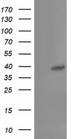
HEK293T cells were transfected with the pCMV6-ENTRY control (Left lane) or pCMV6-ENTRY TBC1D21 (RC206651, Right lane) cDNA for 48 hrs and lysed. Equivalent amounts of cell lysates (5 ug per lane) were separated by SDS-PAGE and immunoblotted with anti-TBC1D21.
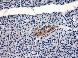
Immunohistochemical staining of paraffin-embedded Human pancreas tissue within the normal limits using anti-TBC1D21 mouse monoclonal antibody. (Heat-induced epitope retrieval by 10mM citric buffer, pH6.0, 100C for 10min, TA503719)
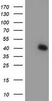
HEK293T cells were transfected with the pCMV6-ENTRY control (Left lane) or pCMV6-ENTRY TBC1D21 (RC206651, Right lane) cDNA for 48 hrs and lysed. Equivalent amounts of cell lysates (5 ug per lane) were separated by SDS-PAGE and immunoblotted with anti-TBC1D21.
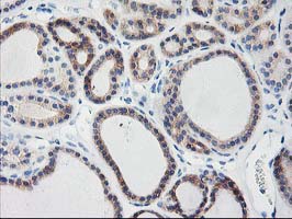
Immunohistochemical staining of paraffin-embedded Carcinoma of Human thyroid tissue using anti-TBC1D21 mouse monoclonal antibody. (Heat-induced epitope retrieval by 10mM citric buffer, pH6.0, 100C for 10min, TA503741)
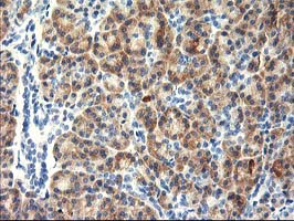
Immunohistochemical staining of paraffin-embedded Human pancreas tissue within the normal limits using anti-TBC1D21 mouse monoclonal antibody. (Heat-induced epitope retrieval by 10mM citric buffer, pH6.0, 100C for 10min, TA503741)
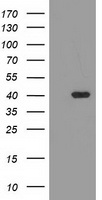
HEK293T cells were transfected with the pCMV6-ENTRY control (Left lane) or pCMV6-ENTRY TBC1D21 (RC206651, Right lane) cDNA for 48 hrs and lysed. Equivalent amounts of cell lysates (5 ug per lane) were separated by SDS-PAGE and immunoblotted with anti-TBC1D21.
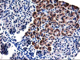
Immunohistochemical staining of paraffin-embedded Human pancreas tissue within the normal limits using anti-TBC1D21 mouse monoclonal antibody. (Heat-induced epitope retrieval by 10mM citric buffer, pH6.0, 100C for 10min, TA503742)
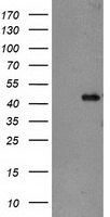
HEK293T cells were transfected with the pCMV6-ENTRY control (Left lane) or pCMV6-ENTRY TBC1D21 (RC206651, Right lane) cDNA for 48 hrs and lysed. Equivalent amounts of cell lysates (5 ug per lane) were separated by SDS-PAGE and immunoblotted with anti-TBC1D21.
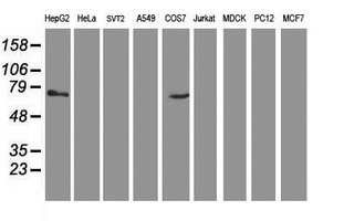
Western blot analysis of extracts (35ug) from 9 different cell lines by using anti-TBC1D21 monoclonal antibody.

HEK293T cells were transfected with the pCMV6-ENTRY control (Left lane) or pCMV6-ENTRY TBC1D21 (RC206651, Right lane) cDNA for 48 hrs and lysed. Equivalent amounts of cell lysates (5 ug per lane) were separated by SDS-PAGE and immunoblotted with anti-TBC1D21.
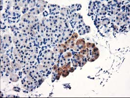
Immunohistochemical staining of paraffin-embedded Human pancreas tissue within the normal limits using anti-TBC1D21 mouse monoclonal antibody. (Heat-induced epitope retrieval by 10mM citric buffer, pH6.0, 100C for 10min, TA503763)
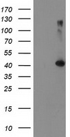
HEK293T cells were transfected with the pCMV6-ENTRY control (Left lane) or pCMV6-ENTRY TBC1D21 (RC206651, Right lane) cDNA for 48 hrs and lysed. Equivalent amounts of cell lysates (5 ug per lane) were separated by SDS-PAGE and immunoblotted with anti-TBC1D21.
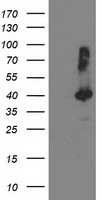
HEK293T cells were transfected with the pCMV6-ENTRY control (Left lane) or pCMV6-ENTRY TBC1D21 (RC206651, Right lane) cDNA for 48 hrs and lysed. Equivalent amounts of cell lysates (5 ug per lane) were separated by SDS-PAGE and immunoblotted with anti-TBC1D21.
- Quick-Filters:
- Highly Validated (2)
Primary Antibodies
| Catalog No. | Host | Iso. | Clone | Pres. | React. | Applications | |
|---|---|---|---|---|---|---|---|
| TA503671 | TBC1D21 antibody |
||||||
| TBC1D21 mouse monoclonal antibody, clone OTI2A6 (formerly 2A6) | |||||||
 |
Mouse | IgG2b | OTI2A6 | Purified | Hu | F, P, WB |
0.1 ml /
€360.00
|
| OriGene Technologies, Inc. | |||||||
 +3 additional images +3 additional images |
Highly validated antibody!
|
||||||
| TA503718 | TBC1D21 antibody |
||||||
| TBC1D21 mouse monoclonal antibody, clone OTI1C7 (formerly 1C7) | |||||||
 |
Mouse | IgG2b | OTI1C7 | Purified | Hu | P, WB |
0.1 ml /
€360.00
|
| OriGene Technologies, Inc. | |||||||
| +3 additional images |
Highly validated antibody!
|
||||||
| TA503710 | TBC1D21 antibody |
||||||
| TBC1D21 mouse monoclonal antibody, clone OTI6F9 (formerly 6F9) | |||||||
 |
Mouse | IgG1 | OTI6F9 | Purified | Hu | P, WB |
0.1 ml /
€360.00
|
| OriGene Technologies, Inc. | |||||||
| +1 additional image |
|
||||||
| TA503716 | TBC1D21 antibody |
||||||
| TBC1D21 mouse monoclonal antibody, clone OTI8B7 (formerly 8B7) | |||||||
 |
Mouse | IgG2b | OTI8B7 | Purified | Hu | WB |
0.1 ml /
€360.00
|
| OriGene Technologies, Inc. | |||||||
|
|||||||
| TA503719 | TBC1D21 antibody |
||||||
| TBC1D21 mouse monoclonal antibody, clone OTI3C2 (formerly 3C2) | |||||||
 |
Mouse | IgG2b | OTI3c2 | Purified | Hu | P, WB |
0.1 ml /
€360.00
|
| OriGene Technologies, Inc. | |||||||
| +1 additional image |
|
||||||
| TA503741 | TBC1D21 antibody |
||||||
| TBC1D21 mouse monoclonal antibody, clone OTI1A8 (formerly 1A8) | |||||||
 |
Mouse | IgG2b | OTI1A8 | Purified | Hu | P, WB |
0.1 ml /
€360.00
|
| OriGene Technologies, Inc. | |||||||
| +2 additional images |
|
||||||
| TA503742 | TBC1D21 antibody |
||||||
| TBC1D21 mouse monoclonal antibody, clone OTI3F1 (formerly 3F1) | |||||||
 |
Mouse | IgG2b | OTI3F1 | Purified | Hu | P, WB |
0.1 ml /
€379.00
|
| OriGene Technologies, Inc. | |||||||
| +2 additional images |
|
||||||
| TA503762 | TBC1D21 antibody |
||||||
| TBC1D21 mouse monoclonal antibody, clone OTI7B1 (formerly 7B1) | |||||||
 |
Mouse | IgG1 | OTI7B1 | Purified | Hu | WB |
0.1 ml /
€360.00
|
| OriGene Technologies, Inc. | |||||||
|
|||||||
| TA503763 | TBC1D21 antibody |
||||||
| TBC1D21 mouse monoclonal antibody, clone OTI7G10 (formerly 7G10) | |||||||
 |
Mouse | IgG1 | OTI7G10 | Purified | Hu | P, WB |
0.1 ml /
€360.00
|
| OriGene Technologies, Inc. | |||||||
| +1 additional image |
|
||||||
| TA503764 | TBC1D21 antibody |
||||||
| TBC1D21 mouse monoclonal antibody, clone OTI8C1 (formerly 8C1) | |||||||
 |
Mouse | IgG2a | OTI8C1 | Purified | Hu | WB |
0.1 ml /
€360.00
|
| OriGene Technologies, Inc. | |||||||
|
|||||||
| AP54166PU-N | TBC1D21 (N-term) antibody |
||||||
 |
Rabbit | Ig | Aff - Purified | Hu | WB |
0.4 ml /
€415.00
|
|
| OriGene Technologies GmbH | |||||||

Lysates
| Catalog No. | ||
|---|---|---|
| LY407069 | TBC1D21 overexpression lysate |
|
 |
0.1 mg /
€295.00
|
|
| OriGene Technologies, Inc. |