TLK1 antibody
Principal name
TLK1 antibody
Alternative names for TLK1 antibody
Tousled-like kinase 1, KIAA0137, Serine/threonine-protein kinase tousled-like 1, PKU-beta
Ncbi ID
Available reactivities
Available hosts
Available applications
Enzyme Immunoassay (E), Western blot / Immunoblot (WB), Paraffin Sections (P), ELISA (detection) (E(detection))
Background of TLK1 antibody
- Kit Component:
- - KN317615G1, Tlk1 gRNA vector 1 in pCas-Guide vector
- KN317615G2, Tlk1 gRNA vector 2 in pCas-Guide vector
- KN317615D, donor vector containing left and right homologous arms and Luciferase-Puro functional cassette
- GE100003, scramble sequence in pCas-Guide vector - CRISPR Manual:
- CRISPR/CAS9 Genome Editing Manual
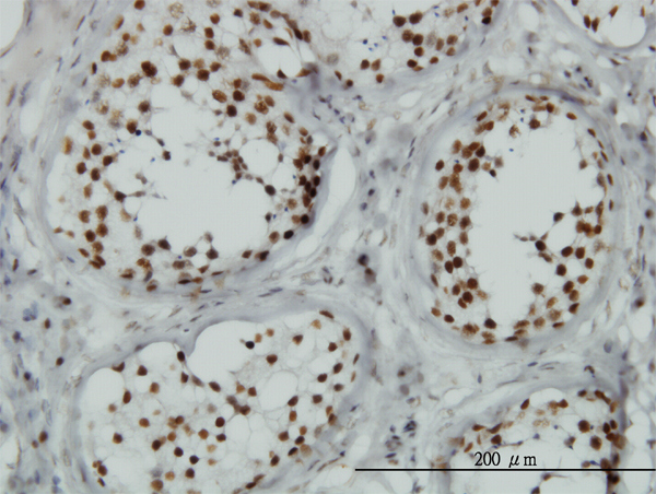
Immunoperoxidase of monoclonal antibody to TLK1 on formalin-fixed paraffin-embedded human testis. [antibody concentration 1 ug/ml]
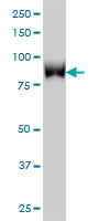
TLK1 monoclonal antibody (M01), clone 4B3 Western Blot analysis of TLK1 expression in HeLa NE ( Cat # L013V3 ).
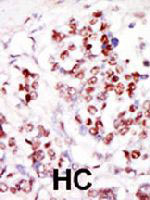
Formalin-fixed and paraffin-embedded human cancer tissue reacted with TLK1 polyclonal antibody ( Cat # PAB3318 ) , which was peroxidase-conjugated to the secondary antibody, followed by DAB staining. This data demonstrates the use of this antibody for immunohistochemistry; clinical relevance has not been evaluated. HC = hepatocarcinoma.

Western blot analysis of TLK1 polyclonal antibody ( Cat # PAB3318 ) in HL-60 cell lysate. TLK1 ( Arrow ) was detected using purified polyclonal antibody. Secondary HRP-anti-rabbit was used for signal visualization with chemiluminescence.
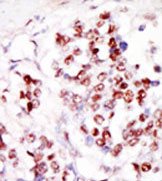
Formalin-fixed and paraffin-embedded human cancer tissue reacted with the primary antibody, which was peroxidase-conjugated to the secondary antibody, followed by DAB staining.
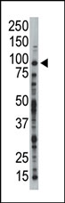
Western blot analysis of anti-TLK1 Pab in HL-60 cell lysate. TLK1 (Arrow) was detected using purified Pab. Secondary HRP-anti-rabbit was used for signal visualization with chemiluminescence.
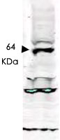
Western blot using TLK1 polyclonal antibody ( Cat # PAB10238 ) shows detection of aband ~ 64 kDa corresponding to isoform 3 of human TLK1 ( arrowhead ).Specific reactivity with this band is blocked when the antibody is pre-incubated with the immunizing peptide ( data not shown ).Approximately 30 µg of a HeLa nuclear extract was separated by SDS-PAGE and transferred onto nitrocellulose.After blocking the membrane was probed with the primary antibody diluted to 1 : 250 for 2h at room temperature followed by washes and reaction with a 1 : 10,000 dilution of IRDyeTM800 conjugated Gt-a-RabbitIgG [H&L] MX for 45 min at room temperature.
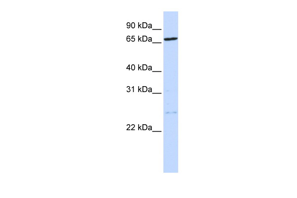
Human 721_B; WB Suggested Anti-TLK1 Antibody Titration: 0.2-1 ug/ml. ELISA Titer: 1:62500. Positive Control: 721_B cell lysate. TLK1 is strongly supported by BioGPS gene expression data to be expressed in Human 721_B cells.; TLK1 antibody - N-terminal region (ARP58340_P050) in Human 721_B cells using Western Blot

Western blot analysis of TLK1 expression in HeLa (A); NIH3T3 (B); PC12 (C) whole cell lysates.
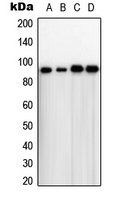
Western blot analysis of TLK1 expression in HeLa (A); A431 (B); NIH3T3 (C); rat thymus (D) whole cell lysates.
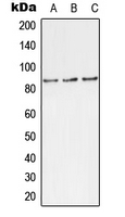
Western blot analysis of TLK1 (pS743) expression in MCF7 (A); rat brain (B); H9C2 (C) whole cell lysates.
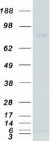
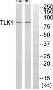
Western blot analysis of extracts from rat brain cells and NIH/3T3 cells, using TLK1 antibody.The lane on the right is treated with the synthesized peptide.
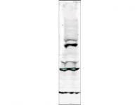
WB using Anti-Tlk-1 antibody shows detection of a band ~64 kDa corresponding to isoform 3 of human Tlk-1 (arrowhead). Specific reactivity with this band is blocked when the antibody is pre-incubated with the immunizing peptide (data not shown). The primary antibody diluted to 1:250 for 2h at RTfollowed by washes and reaction with a 1:10,000 dilution of IRDye 800 conjugated Gt-a-Rabbit IgG [H&L] MX for 45 min at room temperature.
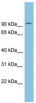
WB Suggested Anti-TLK1 Antibody Titration: 0.2-1 ug/ml; ELISA Titer: 1:312500; Positive Control: ACHN cell lysate
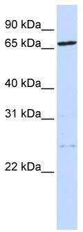
WB Suggested Anti-TLK1 Antibody Titration: 0.2-1 ug/ml; ELISA Titer: 1:62500; Positive Control: 721_B cell lysateTLK1 is strongly supported by BioGPS gene expression data to be expressed in Human 721_B cells
Primary Antibodies
| Catalog No. | Host | Iso. | Clone | Pres. | React. | Applications | |
|---|---|---|---|---|---|---|---|
| TA312355 | TLK1 antibody |
||||||
| Rabbit polyclonal anti-TLK1 antibody | |||||||
 |
Rabbit | IgG | Purified | Hu | E, WB |
0.1 ml /
€335.00
|
|
| OriGene Technologies, Inc. | |||||||
| TA319197 | TLK1 pSer695 antibody |
||||||
| Rabbit polyclonal anti-Tlk1 antibody | |||||||
![WB using Anti-Tlk-1 antibody shows detection of a band ~64 kDa corresponding to isoform 3 of human Tlk-1 (arrowhead). Specific reactivity with this band is blocked when the antibody is pre-incubated with the immunizing peptide (data not shown). The primary antibody diluted to 1:250 for 2h at RTfollowed by washes and reaction with a 1:10,000 dilution of IRDye 800 conjugated Gt-a-Rabbit IgG [H&L] MX for 45 min at room temperature.](https://m1.acris-antibodies.com/catalog/product/cache/1/small_image/50x50/9df78eab33525d08d6e5fb8d27136e95/OriGene/600-401-469-1-W.jpg) |
Rabbit | Aff - Purified | Hu | E |
0.1 mg /
€515.00
|
||
| OriGene Technologies, Inc. | |||||||
| AP15060PU-N | TLK1 (C-term) antibody |
||||||
 |
Rabbit | Ig | Purified | Hu | P, WB |
0.4 ml /
€415.00
|
|
| OriGene Technologies GmbH | |||||||
 +1 additional image +1 additional image |
|||||||
Proteins & Growth Factors
| Catalog No. | Species | Pres. | Purity | Source | |
|---|---|---|---|---|---|
| TP307653 | TLK1 (transcript variant 1) |
||||
| Recombinant protein of human tousled-like kinase 1 (TLK1), transcript variant 1 | |||||
 |
Human | > 80 % Preparation: Recombint protein was captured through anti-DDK affinity column followed by conventiol chromatography steps. Purity Detail: > 80% as determined by SDS-PAGE and Coomassie blue staining. |
HEK293 cells |
20 µg /
€748.00
|
|
| OriGene Technologies, Inc. | |||||
Mouse Lenti ORF Particles
| Catalog No. | ||
|---|---|---|
| MR210263L2V | Lenti ORF particles, Tlk1 (GFP-tagged) - Mouse tousled-like kinase 1 (Tlk1), 200 uL, >10^7 TU/mL |
|
 |
200 µl /
€1,560.00
|
|
| OriGene Technologies, Inc. | ||
| MR210263L1V | Lenti ORF particles, Tlk1 (Myc-DDK-tagged) - Mouse tousled-like kinase 1 (Tlk1), 200 uL, >10^7 TU/mL |
|
|
200 µl /
€1,400.00
|
|
| OriGene Technologies, Inc. | ||