TNK1 antibody
Principal name
TNK1 antibody
Alternative names for TNK1 antibody
Kos1, Non-receptor tyrosine-protein kinase TNK1, CD38 negative kinase 1
Ncbi ID
Available reactivities
Available hosts
Available applications
Western blot / Immunoblot (WB), Enzyme Immunoassay (E), Paraffin Sections (P), Immunocytochemistry/Immunofluorescence (ICC/IF)
Background of TNK1 antibody
- Kit Component:
- - KN318009G1, Tnk1 gRNA vector 1 in pCas-Guide vector
- KN318009G2, Tnk1 gRNA vector 2 in pCas-Guide vector
- KN318009D, donor vector containing left and right homologous arms and Luciferase-Puro functional cassette
- GE100003, scramble sequence in pCas-Guide vector - CRISPR Manual:
- CRISPR/CAS9 Genome Editing Manual
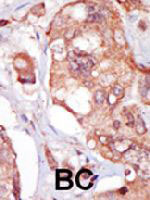
Formalin-fixed and paraffin-embedded human cancer tissue reacted with TNK1 polyclonal antibody ( Cat # PAB3441 ) , which was peroxidase-conjugated to the secondary antibody, followed by AEC staining. This data demonstrates the use of this antibody for immunohistochemistry ; clinical relevance has not been evaluated. BC = breast carcinoma.
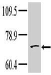
Western blot analysis of TNK1 polyclonal antibody ( Cat # PAB3441 ) in Hela cell line lysate. Dilution of anti-TNK1 was 1 : 100 ; dilution of secondary antibody ( goat anti-rabbit-HRP ) was 1 : 7000. Data and protocol courtesy of Dr. Richard Lu, Partners HealthCare System at Harvard University.

Formalin-fixed and paraffin-embedded human cancer tissue reacted with the primary antibody, which was peroxidase-conjugated to the secondary antibody, followed by AEC staining.
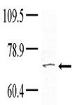
Western blot analysis of anti-TNK1 pab in Hela cell line lysate. Dilution of anti-TNK1 was 1:100; dilution of secondary antibody (goat anti-rabbit-HRP) was 1:7000.
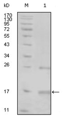
Western blot analysis using TNK1 mouse mAb against truncated TNK1-His recombinant protein (1).
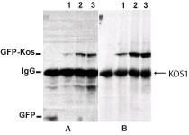
Western Blot: Kos1/Tnk1 Antibody [TA336625] - T47D cells expressing GFP/GFP-Kos were lysed in RIPA lysis buffer. 500ug protein from clarified cell lysates was immunoprecipitated with an anti-GFP antibody. The washed immunoprecipitates were electrophoresed

Immunocytochemistry/Immunofluorescence: Kos1/Tnk1 Antibody [TA336625] - Kos1 staining in Hela cells detected with a Dylight 488 labeled secondary antibody.

Gel: 8%SDS-PAGE, Lysate: 40 ug, Lane: Human fetal brain tissue, Primary antibody: (TNK1 Antibody) at dilution 1/400, Secondary antibody: Goat anti rabbit IgG at 1/8000 dilution, Exposure time: 5 minutes
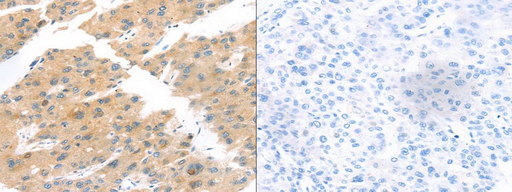
The image on the left is immunohistochemistry of paraffin-embedded Human liver cancer tissue using (TNK1 Antibody) at dilution 1/20, on the right is treated with synthetic peptide. (Original magnification: ×200)
Primary Antibodies
| Catalog No. | Host | Iso. | Clone | Pres. | React. | Applications | |
|---|---|---|---|---|---|---|---|
| TA336625 | TNK1 antibody |
||||||
| Rabbit Polyclonal Kos1/Tnk1 Antibody | |||||||
![Western Blot: Kos1/Tnk1 Antibody [TA336625] - T47D cells expressing GFP/GFP-Kos were lysed in RIPA lysis buffer. 500ug protein from clarified cell lysates was immunoprecipitated with an anti-GFP antibody. The washed immunoprecipitates were electrophoresed](https://m1.acris-antibodies.com/catalog/product/cache/1/small_image/50x50/9df78eab33525d08d6e5fb8d27136e95/OriGene/NB300-237-1-W.jpg) |
Rabbit | Purified | Hu, Ms | ICC/IF, WB |
0.1 ml /
€335.00
|
||
| OriGene Technologies, Inc. | |||||||
 +1 additional image +1 additional image |
|||||||
| TA351832 | TNK1 antibody |
||||||
| Rabbit Polyclonal Anti-TNK1 Antibody | |||||||
 |
Rabbit | IgG | Purified | Hu, Ms | E, P, WB |
0.1 ml /
€335.00
|
|
| OriGene Technologies, Inc. | |||||||
| +1 additional image |
|||||||
| AM06239SU-N | TNK1 antibody |
||||||
 |
Mouse | IgG1 | 1B5G3 | Ascites | Hu | E, WB |
0.1 ml /
€340.00
|
| OriGene Technologies GmbH | |||||||
| AP14482PU-N | TNK1 (Center) antibody |
||||||
 |
Rabbit | Ig | Purified | Hu | P, WB |
0.4 ml /
€415.00
|
|
| OriGene Technologies GmbH | |||||||
| +1 additional image |
|||||||
| RA19032-100 | TNK1 antibody |
||||||
 |
Rabbit | Aff - Purified | Ms | WB |
0.1 mg /
€270.00
|
||
| Neuromics Antibodies | |||||||
Lysates
| Catalog No. | ||
|---|---|---|
| LY401305 | TNK1 overexpression lysate |
|
|
0.1 mg /
€495.00
|
|
| OriGene Technologies, Inc. |
Mouse Lenti ORF Particles
| Catalog No. | ||
|---|---|---|
| MR209890L2V | Lenti ORF particles, Tnk1 (GFP-tagged) - Mouse tyrosine kinase, non-receptor, 1 (Tnk1), 200 uL, >10^7 TU/mL |
|
|
200 µl /
€1,420.00
|
|
| OriGene Technologies, Inc. | ||
| MR209890L1V | Lenti ORF particles, Tnk1 (Myc-DDK-tagged) - Mouse tyrosine kinase, non-receptor, 1 (Tnk1), 200 uL, >10^7 TU/mL |
|
|
200 µl /
€1,270.00
|
|
| OriGene Technologies, Inc. | ||