TUBB1 antibody
Principal name
TUBB1 antibody
Alternative names for TUBB1 antibody
Tubulin beta 1, Tubulin beta-1 chain, beta I Tubulin
Gene ID
81027 (TUBB1)
Ncbi ID
Available reactivities
Available hosts
Available applications
ELISA (detection) (E(detection)), Western blot / Immunoblot (WB), Immunocytochemistry/Immunofluorescence (ICC/IF), Paraffin Sections (P), Frozen Sections (C), Immunoprecipitation (IP)
Background of TUBB1 antibody
- Kit Component:
- - KN207459G1, TUBB1 gRNA vector 1 in pCas-Guide vector
- KN207459G2, TUBB1 gRNA vector 2 in pCas-Guide vector
- KN207459D, donor vector containing left and right homologous arms and Luciferase-Puro functional cassette
- GE100003, scramble sequence in pCas-Guide vector - CRISPR Manual:
- CRISPR/CAS9 Genome Editing Manual

TUBB1 MaxPab polyclonal antibody. Western Blot analysis of TUBB1 expression in rat brain.
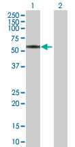
Western Blot analysis of TUBB1 expression in transfected 293T cell line by TUBB1 MaxPab polyclonal antibody.Lane 1: TUBB1 transfected lysate(49.61 KDa).Lane 2: Non-transfected lysate.
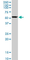
TUBB1 MaxPab rabbit polyclonal antibody. Western Blot analysis of TUBB1 expression in mouse lung.
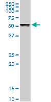
TUBB1 MaxPab rabbit polyclonal antibody. Western Blot analysis of TUBB1 expression in human liver.

Western Blot analysis of TUBB1 expression in transfected 293T cell line (H00081027-T02) by TUBB1 MaxPab polyclonal antibody.Lane 1: TUBB1 transfected lysate(50.30 KDa).Lane 2: Non-transfected lysate.
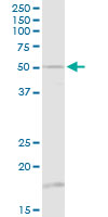
TUBB1 MaxPab rabbit polyclonal antibody. Western Blot analysis of TUBB1 expression in A-431.
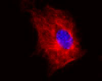
Immunofluorescence staining of NIH/3T3 ( mouse embryonal fibroblast cell line ) using TUBB1 monoclonal antibody, clone TU-06 ( Cat # MAB3656 ) ( detection by Goat anti-mouse IgM Cy®5 ). Nucleus is stained with DAPI ( blue ).
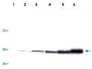
Western Blotting analysis ( reducing conditions ) of HPB-ALL ( human peripheral blood leukemia cell line ).Lane 1 : Negative control.Lane 2, 3, 4, 5, 6 : Immunostaining with TUBB1 monoclonal antibody, clone TU-06 ( Cat # MAB3656 ; dilution 0,5 µg/ml, 1 µg/ml, 2 µg/ml, 4 µg/ml, 5 µg/ml ).
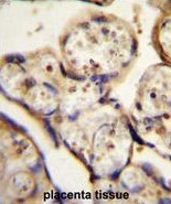
TBB1 Antibody (N-term) (Cat. #AP54162PU-N)immunohistochemistry analysis in formalin fixed and paraffin embedded human placenta tissue followed by peroxidase conjugation of the secondary antibody and DAB staining.This data demonstrates the use of TBB1 Antibody (N-term) for immunohistochemistry. Clinical relevance has not been evaluated.
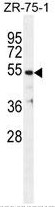
TBB1 Antibody (N-term) (Cat. #AP54162PU-N) western blot analysis in ZR-75-1 cell line lysates (35µg/lane).This demonstrates the TBB1 antibody detected the TBB1 protein (arrow).
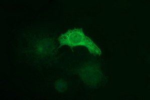
Anti-TUBB1 mouse monoclonal antibody (TA506620) immunofluorescent staining of COS7 cells transiently transfected by pCMV6-ENTRY TUBB1(RC207459).
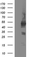
HEK293T cells were transfected with the pCMV6-ENTRY control (Left lane) or pCMV6-ENTRY TUBB1 (RC207459, Right lane) cDNA for 48 hrs and lysed. Equivalent amounts of cell lysates (5 ug per lane) were separated by SDS-PAGE and immunoblotted with anti-TUBB1.
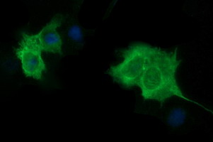
Anti-TUBB1 mouse monoclonal antibody (TA506622) immunofluorescent staining of COS7 cells transiently transfected by pCMV6-ENTRY TUBB1(RC207459).
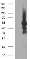
HEK293T cells were transfected with the pCMV6-ENTRY control (Left lane) or pCMV6-ENTRY TUBB1 (RC207459, Right lane) cDNA for 48 hrs and lysed. Equivalent amounts of cell lysates (5 ug per lane) were separated by SDS-PAGE and immunoblotted with anti-TUBB1.
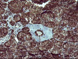
Immunohistochemical staining of paraffin-embedded Human pancreas tissue within the normal limits using anti-TUBB1 mouse monoclonal antibody. (TA506654)
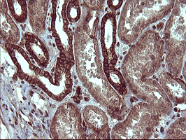
Immunohistochemical staining of paraffin-embedded Human Kidney tissue within the normal limits using anti-TUBB1 mouse monoclonal antibody. (TA506654)
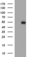
HEK293T cells were transfected with the pCMV6-ENTRY control (Left lane) or pCMV6-ENTRY TUBB1 (RC207459, Right lane) cDNA for 48 hrs and lysed. Equivalent amounts of cell lysates (5 ug per lane) were separated by SDS-PAGE and immunoblotted with anti-TUBB1.
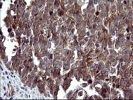
Immunohistochemical staining of paraffin-embedded Adenocarcinoma of Human colon tissue using anti-TUBB1 mouse monoclonal antibody. (Heat-induced epitope retrieval by 10mM citric buffer, pH6.0, 120C for 3min, TA506805)

Immunohistochemical staining of paraffin-embedded Human endometrium tissue within the normal limits using anti-TUBB1 mouse monoclonal antibody. (Heat-induced epitope retrieval by 10mM citric buffer, pH6.0, 120C for 3min, TA506805)
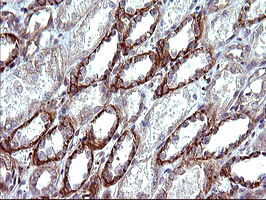
Immunohistochemical staining of paraffin-embedded Human Kidney tissue within the normal limits using anti-TUBB1 mouse monoclonal antibody. (Heat-induced epitope retrieval by 10mM citric buffer, pH6.0, 120C for 3min, TA506805)
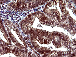
Immunohistochemical staining of paraffin-embedded Adenocarcinoma of Human endometrium tissue using anti-TUBB1 mouse monoclonal antibody. (Heat-induced epitope retrieval by 10mM citric buffer, pH6.0, 120C for 3min, TA506805)
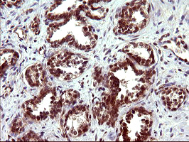
Immunohistochemical staining of paraffin-embedded Carcinoma of Human prostate tissue using anti-TUBB1 mouse monoclonal antibody. (Heat-induced epitope retrieval by 10mM citric buffer, pH6.0, 120C for 3min, TA506805)
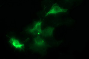
Anti-TUBB1 mouse monoclonal antibody (TA506805) immunofluorescent staining of COS7 cells transiently transfected by pCMV6-ENTRY TUBB1(RC207459).
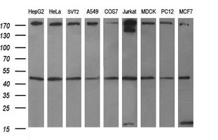
Western blot analysis of extracts (35ug) from 9 different cell lines by using anti-TUBB1 monoclonal antibody (HepG2: human; HeLa: human; SVT2: mouse; A549: human; COS7: monkey; Jurkat: human; MDCK: canine; PC12: rat; MCF7: human).
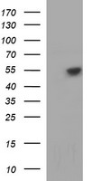
HEK293T cells were transfected with the pCMV6-ENTRY control (Left lane) or pCMV6-ENTRY TUBB1 (RC207459, Right lane) cDNA for 48 hrs and lysed. Equivalent amounts of cell lysates (5 ug per lane) were separated by SDS-PAGE and immunoblotted with anti-TUBB1.
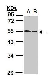
Sample(30 ug whole cell lysate). A:A431. B:Hep G2 . 10% SDS PAGE. TA307997 diluted at 1:1000
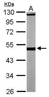
Sample (30 ug of whole cell lysate). A: NIH-3T3. 10% SDS PAGE. TA307997 diluted at 1:1000.
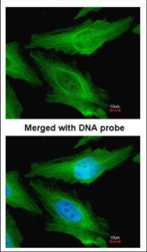
Immunofluorescence analysis of paraformaldehyde-fixed HeLa, using TUBB1(TA307997) antibody at 1:200 dilution.
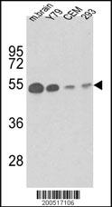
Western blot analysis of anti-TBB1 Antibody (Cat. #TA324323) in mouse brain tissue, Y79, CEM and 293 lysates (35ug/lane). TBB1 (arrow) was detected using the purified Mab.(1ug/ml)
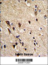
Formalin-fixed and paraffin-embedded human brain reacted with TBB1 Antibody, which was peroxidase-conjugated to the secondary antibody, followed by DAB staining. This data demonstrates the use of this antibody for immunohistochemistry; clinical relevance has not been evaluated.
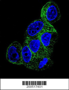
Confocal immunofluorescent analysis of TBB1 Antibody (Cat#TA324323) with HepG2 cell followed by Alexa Fluor® 488-conjugated goat anti-mouse lgG (green). DAPI was used to stain the cell nuclear (blue).
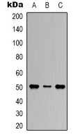
Western blot analysis of Beta1-tubulin expression in Hela (A); mouse brain (B); rat brain (C) whole cell lysates.
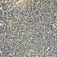
Immunohistochemical analysis of Beta1-tubulin staining in human tonsil formalin fixed paraffin embedded tissue section. The section was pre-treated using heat mediated antigen retrieval with sodium citrate buffer (pH 6.0). The section was then incubated with the antibody at room temperature and detected using an HRP conjugated compact polymer system. DAB was used as the chromogen. The section was then counterstained with haematoxylin and mounted with DPX.
- Quick-Filters:
- Highly Validated (1)
Primary Antibodies
| Catalog No. | Host | Iso. | Clone | Pres. | React. | Applications | |
|---|---|---|---|---|---|---|---|
| TA506805 | TUBB1 antibody |
||||||
| TUBB1 mouse monoclonal antibody, clone OTI4A3 (formerly 4A3) | |||||||
 |
Mouse | IgG2a | OTI4A3 | Purified | Can, Hu, Mky, Ms, Rt | ICC/IF, P, WB |
0.1 ml /
€360.00
|
| OriGene Technologies, Inc. | |||||||
 +7 additional images +7 additional images |
Highly validated antibody!
|
||||||
| TA307997 | TUBB1 antibody |
||||||
| Rabbit Polyclonal antibody to beta Tubulin (tubulin, beta 1) | |||||||
 |
Rabbit | IgG | Aff - Purified | Hu, Ms | ICC/IF, WB |
0.1 ml /
€415.00
|
|
| OriGene Technologies, Inc. | |||||||
| +2 additional images |
|||||||
| TA324323 | TUBB1 antibody |
||||||
| Mouse monoclonal TBB1 Antibody | |||||||
 |
Mouse | IgG1 | 86CT4.4.4 | Purified | Hu, Ms | ICC/IF, P, WB |
0.4 ml /
€415.00
|
| OriGene Technologies, Inc. | |||||||
| +2 additional images |
|||||||
| TA506620 | TUBB1 antibody |
||||||
| TUBB1 mouse monoclonal antibody, clone OTI1D5 (formerly 1D5) | |||||||
 |
Mouse | IgG2a | OTI1D5 | Purified | Hu | ICC/IF, WB |
0.1 ml /
€360.00
|
| OriGene Technologies, Inc. | |||||||
| +1 additional image |
|
||||||
| TA506622 | TUBB1 antibody |
||||||
| TUBB1 mouse monoclonal antibody, clone OTIG5 (formerly G5) | |||||||
 |
Mouse | IgG2a | OTIG5 | Purified | Hu | ICC/IF, WB |
0.1 ml /
€360.00
|
| OriGene Technologies, Inc. | |||||||
| +1 additional image |
|
||||||
| TA506654 | TUBB1 antibody |
||||||
| TUBB1 mouse monoclonal antibody, clone OTI1B9 (formerly 1B9) | |||||||
 |
Mouse | IgG2a | OTI1B9 | Purified | Hu | P, WB |
0.1 ml /
€360.00
|
| OriGene Technologies, Inc. | |||||||
| +2 additional images |
|
||||||
| AP54162PU-N | TUBB1 (N-term) antibody |
||||||
 |
Rabbit | Ig | Aff - Purified | Hu | WB |
0.4 ml /
€415.00
|
|
| OriGene Technologies GmbH | |||||||
| +1 additional image |
|||||||

Lysates
| Catalog No. | ||
|---|---|---|
| LY410723 | TUBB1 overexpression lysate |
|
 |
0.1 mg /
€295.00
|
|
| OriGene Technologies, Inc. |