ACOX1 antibody
Principal name
ACOX1 antibody
Alternative names for ACOX1 antibody
AOX, Palmitoyl-CoA oxidase, Straight-chain acyl-CoA oxidase, Peroxisomal acyl-coenzyme A oxidase 1
Ncbi ID
9606, NP_001171968.1, NM_001185039, 10090, NM_015729, NM_017340, NM_004035, NM_007292
Available reactivities
Available hosts
Available applications
Enzyme Immunoassay (E), Western blot / Immunoblot (WB), ELISA (detection) (E(detection)), Immunocytochemistry/Immunofluorescence (ICC/IF), Immunoprecipitation (IP), Paraffin Sections (P)
Background of ACOX1 antibody
ACOX1 is the first enzyme of the fatty acid beta-oxidation pathway, which catalyzes the desaturation of acyl-CoAs to 2-trans-enoyl-CoAs. It donates electrons directly to molecular oxygen, thereby producing hydrogen peroxide. Defects in this gene result in pseudoneonatal adrenoleukodystrophy, a disease that is characterized by accumulation of very long chain fatty acids.
Function: Catalyzes the desaturation of acyl-CoAs to 2-trans- enoyl-CoAs. Isoform 1 shows highest activity against medium-chain fatty acyl-CoAs and activity decreases with increasing chain length. Isoform 2 is active against a much broader range of substrates and shows activity towards very long-chain acyl-CoAs. Isoform 2 is twice as active as isoform 1 against 16-hydroxy- palmitoyl-CoA and is 25% more active against 1,16-hexadecanodioyl- CoA.
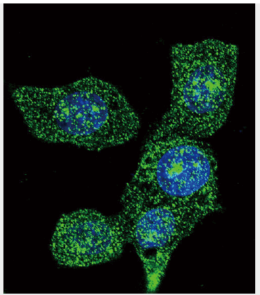
Cellular Location: Peroxisome.
Tissue Location: Widely expressed with highest levels of isoform 1 and isoform 2 detected in testis. Isoform 1 is expressed at higher levels than isoform 2 in liver and kidney while isoform 2 levels are higher in brain, lung, muscle, white adipose tissue and testis. Levels are almost equal in heart.
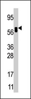
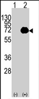
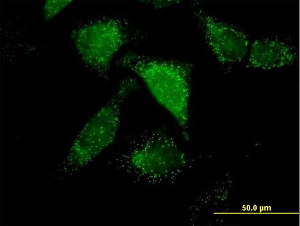
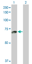
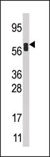
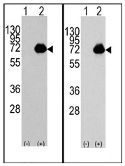
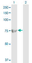
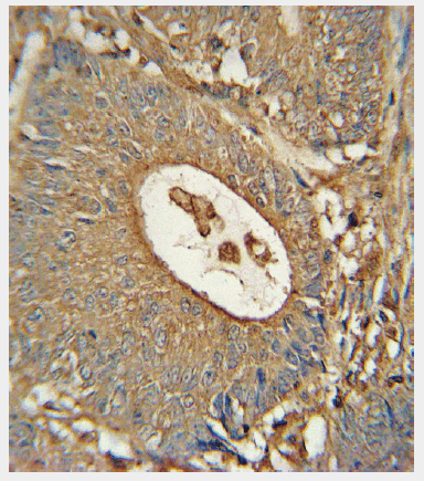

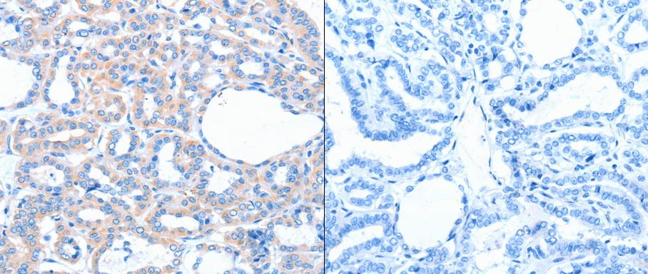
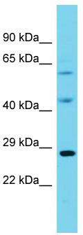
Primary Antibodies
| Catalog No. | Host | Iso. | Clone | Pres. | React. | Applications | |
|---|---|---|---|---|---|---|---|
| TA324047 | ACOX1 antibody |
||||||
| Anti-ACOX1 rabbit polyclonal antibody | |||||||
 |
Rabbit | IgG | Purified | Hu, Ms, Rt | E, P, WB |
0.1 ml /
€335.00
|
|
| OriGene Technologies, Inc. | |||||||
 +1 additional image +1 additional image |
|||||||
| TA324048 | ACOX1 antibody |
||||||
| Anti-ACOX1 Rabbit Polyclonal Antibody | |||||||
 |
Rabbit | IgG | Purified | Hu, Ms, Rt | E, P |
0.1 ml /
€335.00
|
|
| OriGene Technologies, Inc. | |||||||
| +1 additional image |
|||||||