Bax antibody
Principal name
Bax antibody
Alternative names for Bax antibody
Apoptosis regulator BAX, BCL2L4, Bcl2-L-4, Bcl-2-like protein 4, BCL2-associated X protein
Available reactivities
Available hosts
Available applications
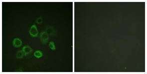
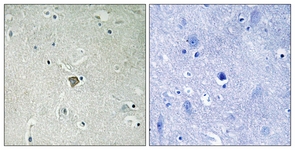
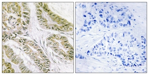
Immunoprecipitation (IP), Western blot / Immunoblot (WB), Paraffin Sections (P), Enzyme Immunoassay (E), Immunocytochemistry/Immunofluorescence (ICC/IF), Frozen Sections (C), Flow Cytometry (F)
Background of Bax antibody
The human protein Bax sits at a critical regulatory junction of apoptosis, or programmed cell death. Activated Bax changes conformation, inserts into the MOM (Mitochondrial Outer Membrane), oligomerizes, and induces MOM permeabilization, causing the release of cytochrome c, which effectively commits the cell to die. (Ma J et al., 2012). Mechanisms of membrane perforation include formation of hetero-oligomeric complexes of Bax with other pro-apoptotic proteins such as Bak, or formation of lipidic pores physically aided by mitochondrial membrane-inserted proteins (Garg P et al., 2012). Connexins play important roles in many physiological and pathological processes. In the context of apoptosis, Cx43 translocated to the mitochondria, where it interacted with Bax to initiate the mitochondrial apoptotic pathway. The 241-382 aa region of Cx43 was required for interaction with Bax. Furthermore, this region was responsible for permeabilizing mitochondrial membrane potential. Recent studies elucidate a novel mechanism of the Cx43-mediated regulation of apoptosis in pancreatic cancer (Sun Y et al., 2012).
Bax acts as a biomarker that exhibited a difference in sub-cellular localization between normal OCSE (Oral Cavity Squamous Epithelium) and OSCC (Oral Cavity Squamous Cell Carcinoma) and was also the only apoptotic protein significantly associated with prognosis. The translocation of Bax from the nucleus to the cytoplasm in OSCC is consistent with increased Bax function at the mitochondria, leading to improved sensitivity to radiotherapy-induced apoptosis in tumours with elevated Bax expression. Bax antibody can be used to study the intracellular redistribution of Bax protein upon induction of apoptosis and its unique subcellular localization. This product can also be used in immunoblot analysis to estimate variations in the expression of specific proteins involved in apoptosis signaling (Bose P et al., 2012).
General readings
1. Dinh C et al. Short Interfering RNA against Bax Attenuates TNFa-Induced Ototoxicity in Rat Organ of Corti Explants. Otolaryngol Head Neck Surg 148:834-40 (2013). ICC/IF ; Rat .
2. Monsalve DM et al. Human VRK2 modulates apoptosis by interaction with Bcl-xL and regulation of BAX gene expression. Cell Death Dis 4:e513 (2013). Human .
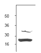
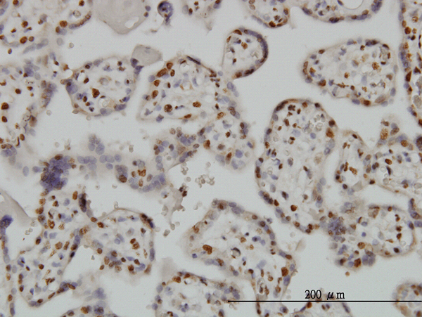
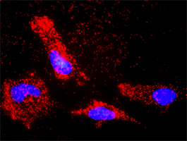
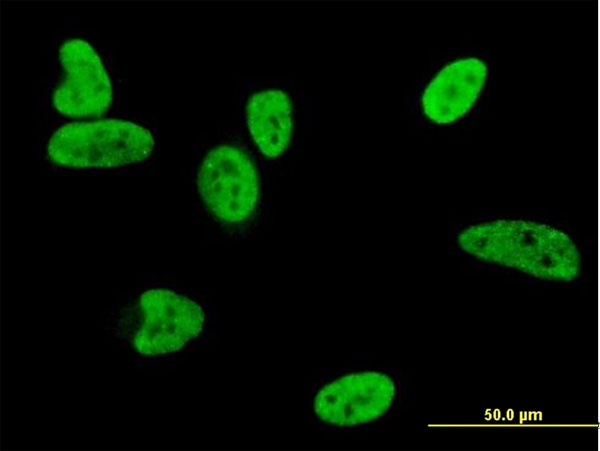

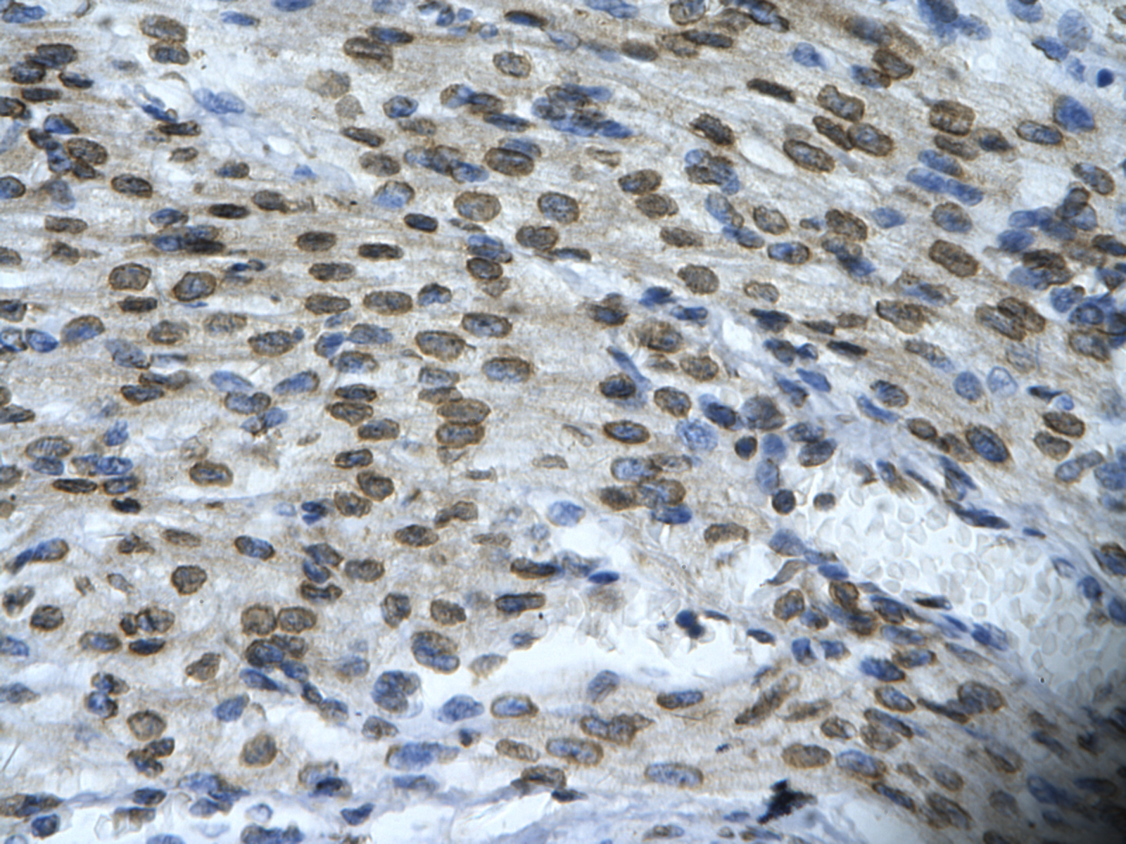
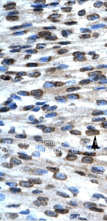
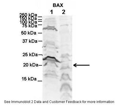
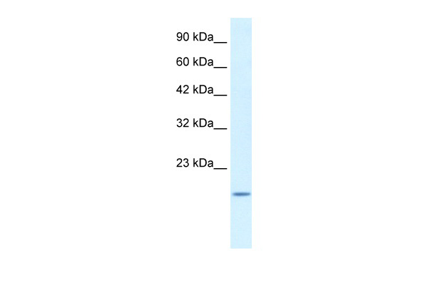
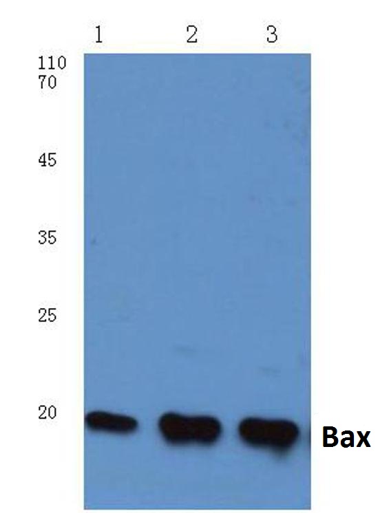
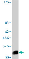
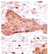
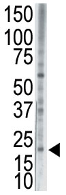
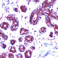
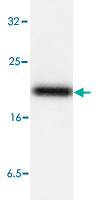
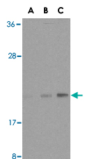
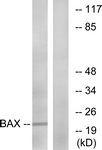
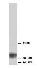
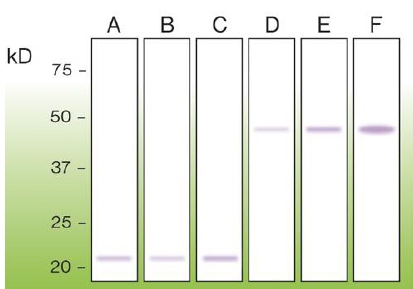
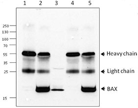
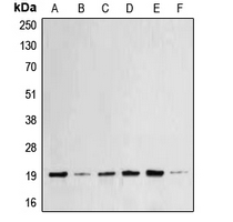
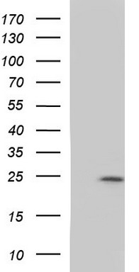
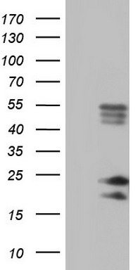
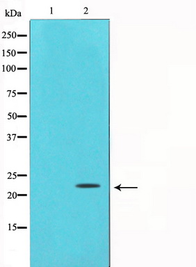
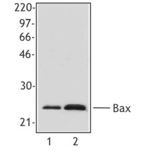
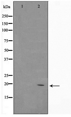
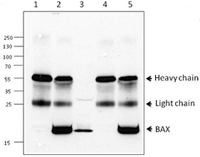
Lane1:MCF-7 cell lysate
Lane2:Raw264.7 cell lysate
Lane3:Rat brain tissue lysate





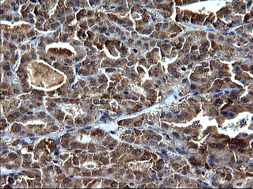
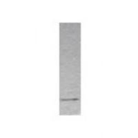
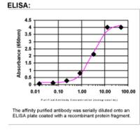

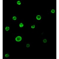

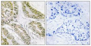
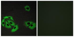
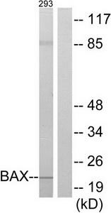
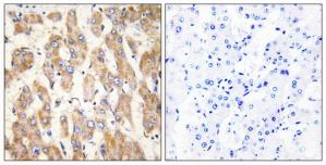
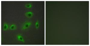
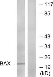

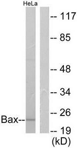
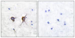
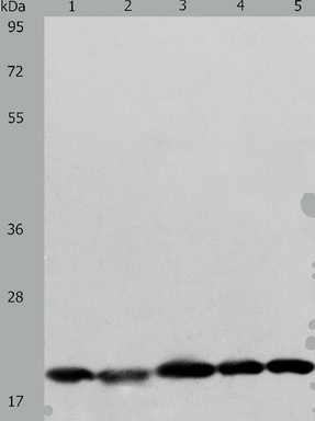
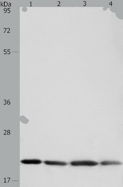
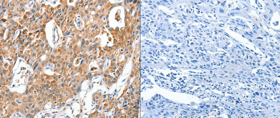

- Quick-Filters:
- Highly Validated (1)

Primary Antibodies
| Catalog No. | Host | Iso. | Clone | Pres. | React. | Applications | |
|---|---|---|---|---|---|---|---|
| TA590174 | Bax antibody |
||||||
| Rabbit Polyclonal Bax Antibody | |||||||
 |
Rabbit | IgG | Purified | Hu | E, P, WB |
0.1 mg /
€409.00
|
|
| OriGene Technologies, Inc. | |||||||
 +4 additional images +4 additional images |
Highly validated antibody! |
||||||
| TA306176 | Bax (N-term) antibody |
||||||
| Rabbit Polyclonal Bax Antibody | |||||||
 |
Rabbit | IgG | Purified | Hu | E, ICC/IF, WB |
0.1 mg /
€415.00
|
|
| OriGene Technologies, Inc. | |||||||
| +2 additional images |
|||||||
| TA312594 | Bax antibody |
||||||
| Rabbit polyclonal anti-BAX antibody | |||||||
 |
Rabbit | IgG | Purified | Hu, Ms | E, P |
0.1 ml /
€335.00
|
|
| OriGene Technologies, Inc. | |||||||
| TA312595 | Bax antibody |
||||||
| Rabbit polyclonal anti-BAX antibody | |||||||
 |
Rabbit | IgG | Purified | Hu, Ms | E, ICC/IF, P |
0.1 ml /
€335.00
|
|
| OriGene Technologies, Inc. | |||||||
| +1 additional image |
|||||||
| TA312596 | Bax antibody |
||||||
| Rabbit polyclonal anti-BAX antibody | |||||||
 |
Rabbit | IgG | Purified | Hu, Ms, Rt | E, ICC/IF, P, WB |
0.1 ml /
€335.00
|
|
| OriGene Technologies, Inc. | |||||||
| +2 additional images |
|||||||
| TA312598 | Bax antibody |
||||||
| Rabbit polyclonal Bax (Ab-167) antibody | |||||||
 |
Rabbit | IgG | Purified | Hu, Ms | E, ICC/IF, WB |
0.1 ml /
€335.00
|
|
| OriGene Technologies, Inc. | |||||||
| +1 additional image |
|||||||
| TA312599 | Bax antibody |
||||||
| Rabbit polyclonal Bax (Thr167) antibody(Phospho-specific) | |||||||
 |
Rabbit | IgG | Purified | Hu, Ms | E, ICC/IF |
0.1 ml /
€335.00
|
|
| OriGene Technologies, Inc. | |||||||
| TA312600 | Bax antibody |
||||||
| Rabbit polyclonal anti-Bax antibody | |||||||
 |
Rabbit | IgG | Purified | Hu, Rt | E, P, WB |
0.1 ml /
€335.00
|
|
| OriGene Technologies, Inc. | |||||||
| +1 additional image |
|||||||
| TA321641 | Bax antibody |
||||||
| Anti-BAX Rabbit Polyclonal Antibody | |||||||
 |
Rabbit | IgG | Purified | Hu, Ms, Rt | E, WB |
0.1 ml /
€335.00
|
|
| OriGene Technologies, Inc. | |||||||
| TA321642 | Bax antibody |
||||||
| Anti-BAX Rabbit Polyclonal Antibody | |||||||
 |
Rabbit | IgG | Purified | Hu, Ms, Rt | E, P, WB |
0.1 ml /
€335.00
|
|
| OriGene Technologies, Inc. | |||||||
| +1 additional image |
|||||||
| TA325255 | Bax antibody |
||||||
| Rabbit Polyclonal Bax Antibody | |||||||
 |
Rabbit | IgG | Purified | Hu, Ms, Rt | P, WB |
0.1 ml /
€335.00
|
|
| OriGene Technologies, Inc. | |||||||
| +1 additional image |
|||||||
| TA326770 | Bax antibody |
||||||
| Bax Rabbit Polyclonal Antibody | |||||||
 |
Rabbit | IgG | Purified | Hu, Ms, Rt |
0.1 mg /
€335.00
|
||
| OriGene Technologies, Inc. | |||||||
| +1 additional image |
|||||||
| TA347321 | Bax antibody |
||||||
| Rabbit Polyclonal Anti-Bax Antibody | |||||||
 |
Rabbit | IgG | Purified | Hu, Ms, Rt | WB |
0.1 mg /
€335.00
|
|
| OriGene Technologies, Inc. | |||||||
| TA890001 | Bax antibody |
||||||
| Rabbit polyclonal BAX Antibody | |||||||
 |
Rabbit | IgG | Purified | Hu | P, WB |
0.1 ml /
€345.00
|
|
| OriGene Technologies, Inc. | |||||||
| +2 additional images |
|||||||
| AP05118PU-N | Bax (beta isoform) antibody |
||||||
 |
Sheep | IgG | Purified | Ms | IP |
0.25 mg /
€420.00
|
|
| OriGene Technologies GmbH | |||||||
| AP06027PU-N | Bax antibody |
||||||
 |
Rabbit | Aff - Purified | Hu, Ms, Rt | IP, P, WB |
0.1 mg /
€370.00
|
||
| OriGene Technologies GmbH | |||||||
| +2 additional images |
|||||||
| AP07277PU-N | Bax antibody |
||||||
 |
Rabbit | Purified | Hu | ICC/IF, P, WB |
50 µg /
€425.00
|
||
| OriGene Technologies GmbH | |||||||
| AP11294PU-N | Bax (BH3 Domain) antibody |
||||||
 |
Rabbit | Ig | Purified | Hu, Ms | P, WB |
0.4 ml /
€415.00
|
|
| OriGene Technologies GmbH | |||||||
| +1 additional image |
|||||||
| AP21052PU-N | Bax antibody |
||||||
 |
Rabbit | Aff - Purified | Hu, Ms, Rt | ICC/IF, WB |
0.1 mg /
€370.00
|
||
| OriGene Technologies GmbH | |||||||
| AP23289PU-N | Bax (N-term) antibody |
||||||
 |
Rabbit | IgG | Aff - Purified | Hu, Ms, Rb, Rt | WB |
0.1 mg /
€390.00
|
|
| OriGene Technologies GmbH | |||||||

