Galectin-3 antibody
Principal name
Galectin-3 antibody
Alternative names for Galectin-3 antibody
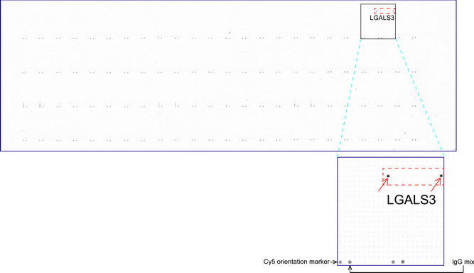
Mac-2, Lgals3, GAL3, GALBP, CBP35, L-31, Galactose-specific lectin 3, IgE-binding protein, 35 kDa lectin, Carbohydrate-binding protein 35, Laminin-binding protein, Lectin L-29, L-34 galactoside-binding lectin
Ncbi ID
NP_002297.2, NP_002297, 9606, NP_034835.1, 10090, AAH01120, NM_001177388
Available reactivities
Available hosts
Available applications
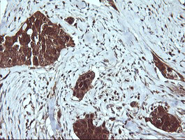
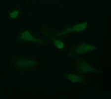
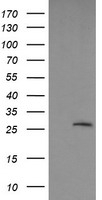
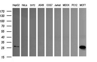
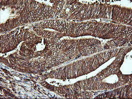
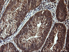
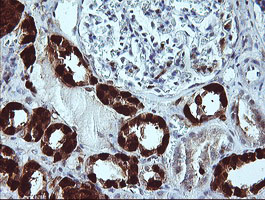
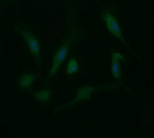
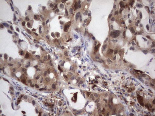
Western blot / Immunoblot (WB), Paraffin Sections (P), Enzyme Immunoassay (E), Functional assay (FN), Frozen Sections (C), Immunocytochemistry/Immunofluorescence (ICC/IF), Flow Cytometry (F), ELISA (detection) (E(detection)), Immunoprecipitation (IP), Luminex (LMNX)
Background of Galectin-3 antibody
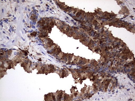
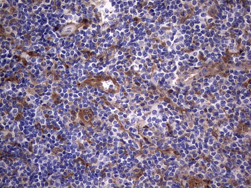
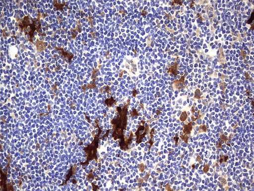
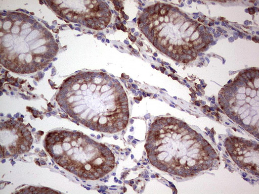
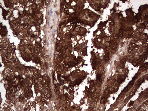
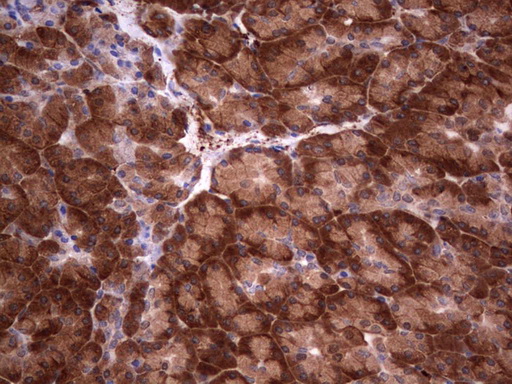
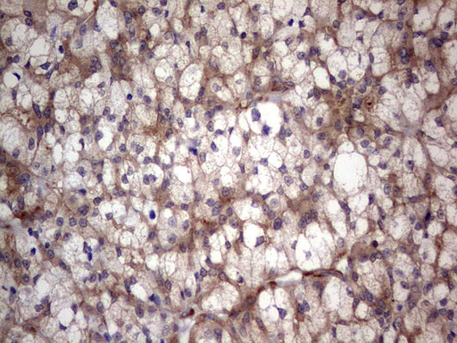
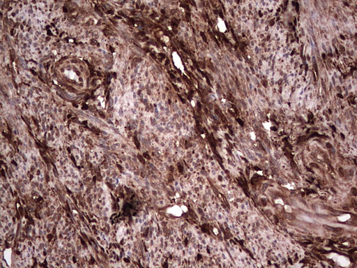
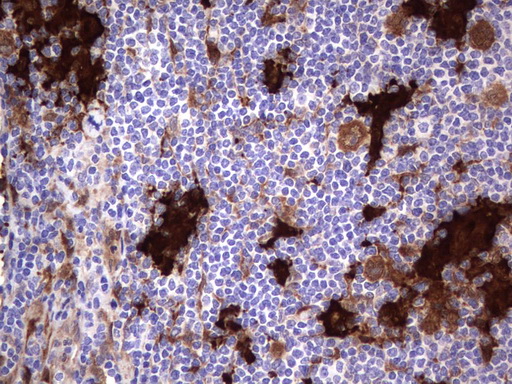
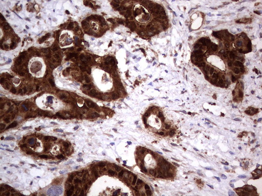
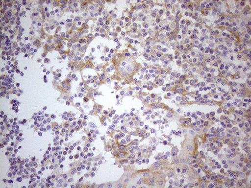
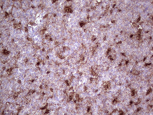
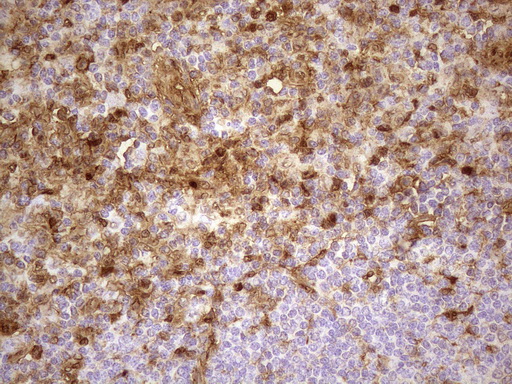
Galectin-3 is a member of the galectin family. The protein is composed of three domains: a small amino-terminal domain, a carboxyl-terminal carbohydrate recognition domain (CRD) and amino-terminal domain containing repeating elements. Galectin-3 is normally distributed in epithelia of many organs and various inflammatory cells, including macrophages, as well as dendritic cells and Kupffer cells. The expression of this lectin is up-regulated during inflammation, cell proliferation, cell differentiation and through trans-activation by viral proteins. The expression is also affected by neoplastic transformation: up-regulated in certain types of lymphomas and thyroid carcinoma, while down-regulated in other types of malignancies, such as colon, breast, ovarian and uterine carcinomas.
Galectin-3 has been shown to function through both intracellular and extracellular actions. Related to its intracellular functions, galectin-3 has been identified as a component of heterogeneous nuclear ribonuclear protein (hnRNP), a factor in pre-mRNA splicing, and has been found to control cell cycle and prevent T cell apoptosis. On the other hand, this protein has also been demonstrated to function as extracellular molecule in activating various types of cells, including monocytes/macrophages, mast cells, neutrophils and lymphocytes. Galectin-3 has been shown to mediate cell-cell and cell-extracellular matrix interactions.
General readings
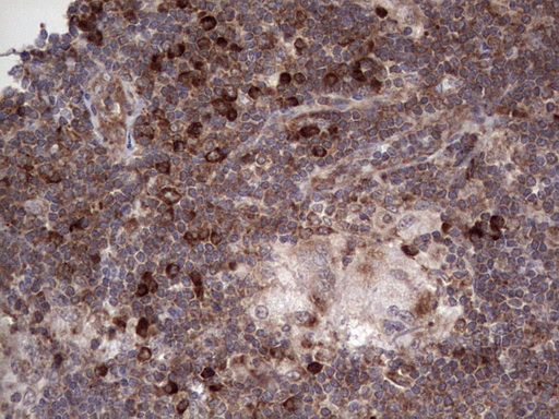
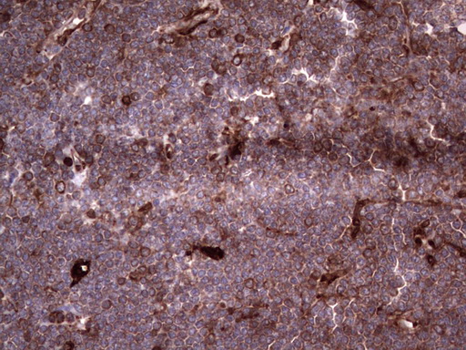
1. Ma F et al. The requirement of CD8+ T cells to initiate and augment acute cardiac inflammatory response to high blood pressure. J Immunol 192:3365-73 (2014). IHC-P,ICC/IF ; Mouse .PubMed: 24600037
2. Lee YJ et al. Spatial and temporal expression, and statin responsiveness of galectin-1 and galectin-3 in murine atherosclerosis. Korean Circ J 43:223-30 (2013). IHC-FoFr ; Mouse .PubMed: 23682281
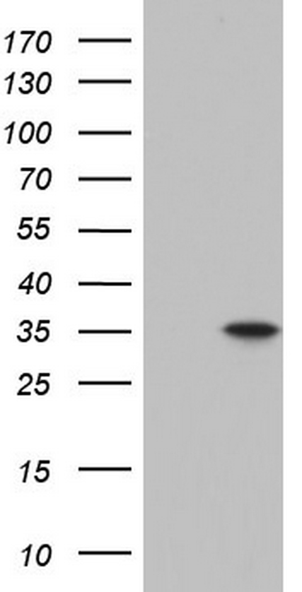
3. Reticker-Flynn NE et al. A combinatorial extracellular matrix platform identifies cell-extracellular matrix interactions that correlate with metastasis. Nat Commun 3:1122 (2012).WB .PubMed: 23047680
4. Doe JA et al. Transgenic overexpression of the a7 integrin reduces muscle pathology and improves viability in the dy(W) mouse model of merosin-deficient congenital muscular dystrophy type 1A. J Cell Sci 124:2287-97 (2011). WB, IHC-Fr ; Mouse .PubMed: 21652631
5. Checinska A et al. Comparative proteomics analysis of caspase-9-protein complexes in untreated and cytochrome c/dATP stimulated lysates of NSCLC cells. J Proteomics 72:575-85 (2009).PubMed: 19118655

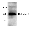
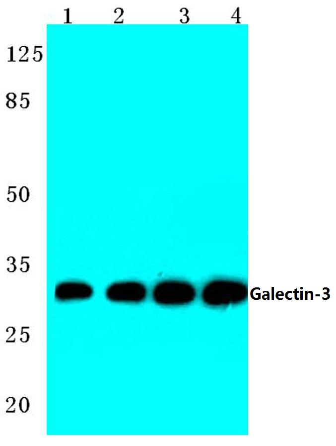
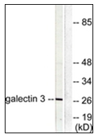
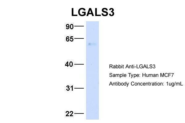
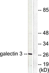
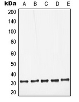
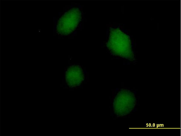
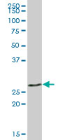
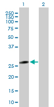
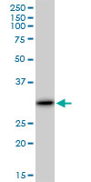
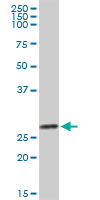
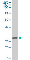
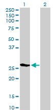
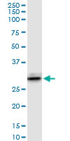
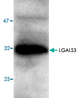
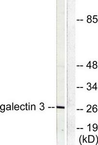
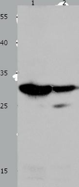
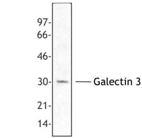
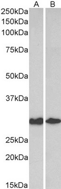
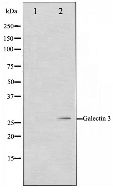
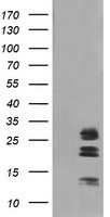
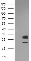
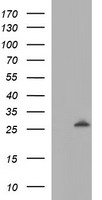
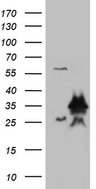
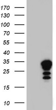
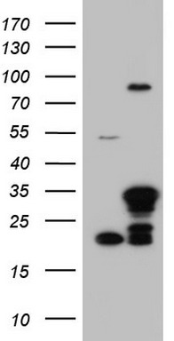
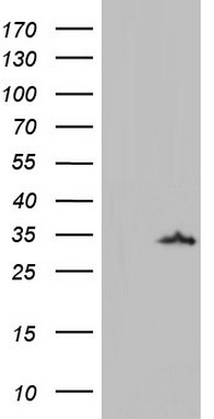
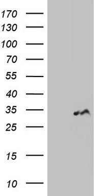
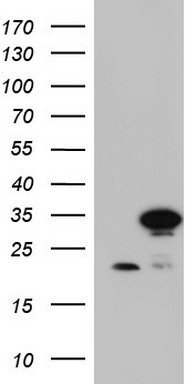
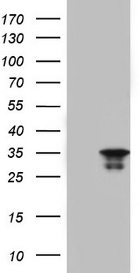
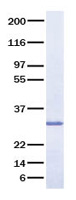
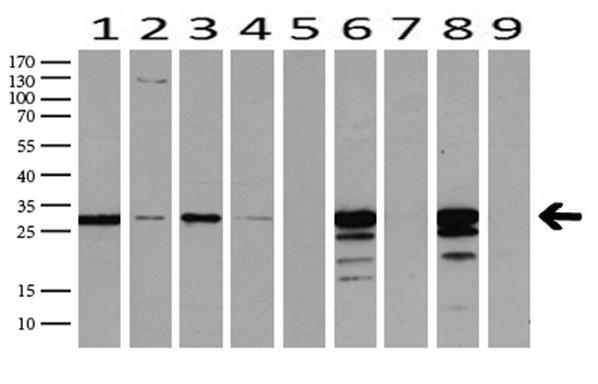
Lane 1: Hela whole cell lysate.
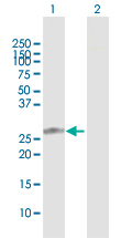
Lane 2: Mouse liver tissue lysate.
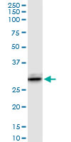
Lane 3: Raw264.7 whole cell lysate.
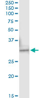
Lane 4: Rat liver tissue lysate.










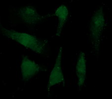
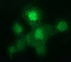

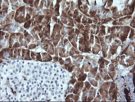




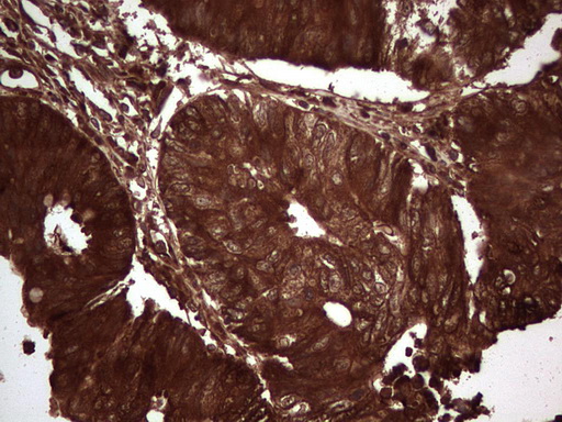

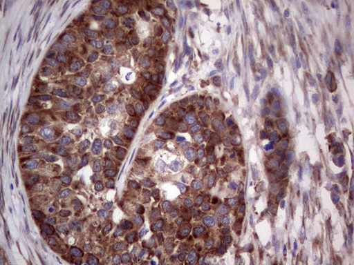
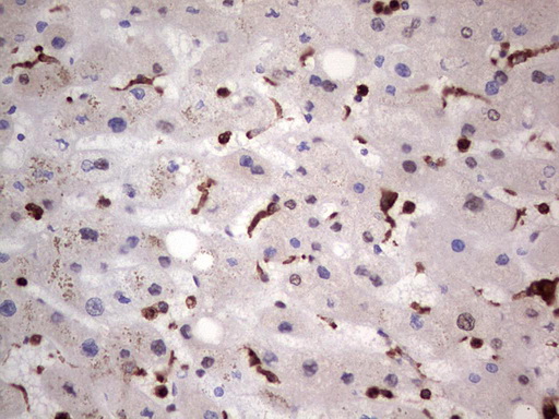
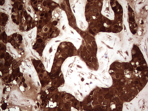




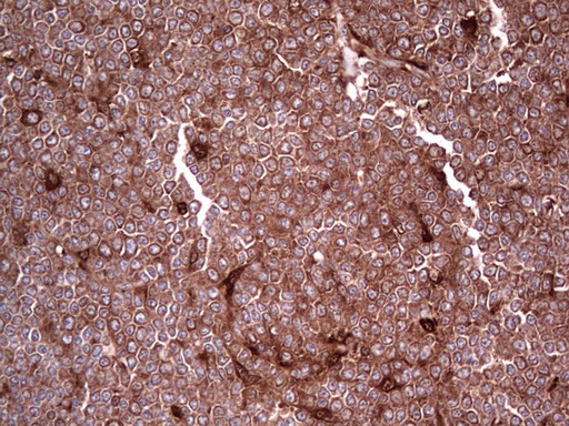
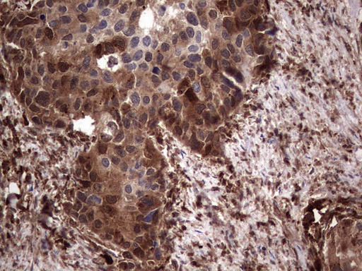
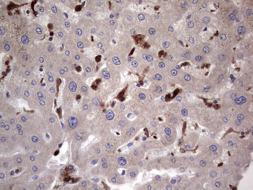
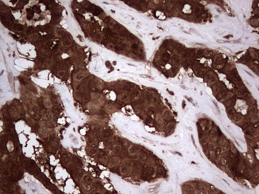
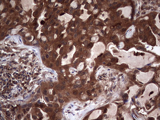
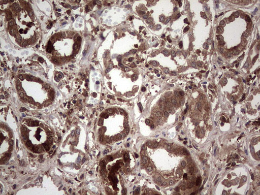
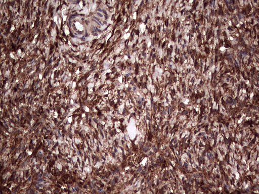







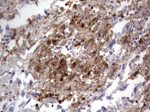
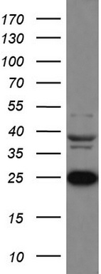

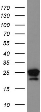
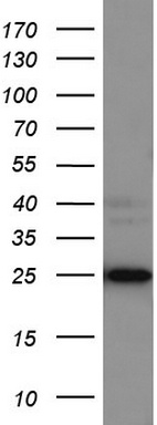
Primary Antibodies
| Catalog No. | Host | Iso. | Clone | Pres. | React. | Applications | |
|---|---|---|---|---|---|---|---|
| AP54962SU-N | Galectin-3 antibody |
||||||
 |
Rabbit | Serum | Hu | C, E, WB |
0.2 ml /
€430.00
|
||
| OriGene Technologies GmbH | |||||||
 +1 additional image +1 additional image |
|||||||

